


Summary: Bright light retinal damage in rodents remains one of the most widely used models mimicking human retinal degenerations such as retinitis pigmentosa (RP) and age-related macular degeneration (AMD).
Model Description
Retinal damage induced by bright light in laboratory rodents has remained one of the most widely used models mimicking human retinal degenerations such as retinitis pigmentosa (RP) and age-related macular degeneration (AMD) for more than five decades.
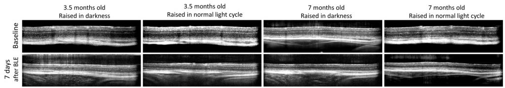
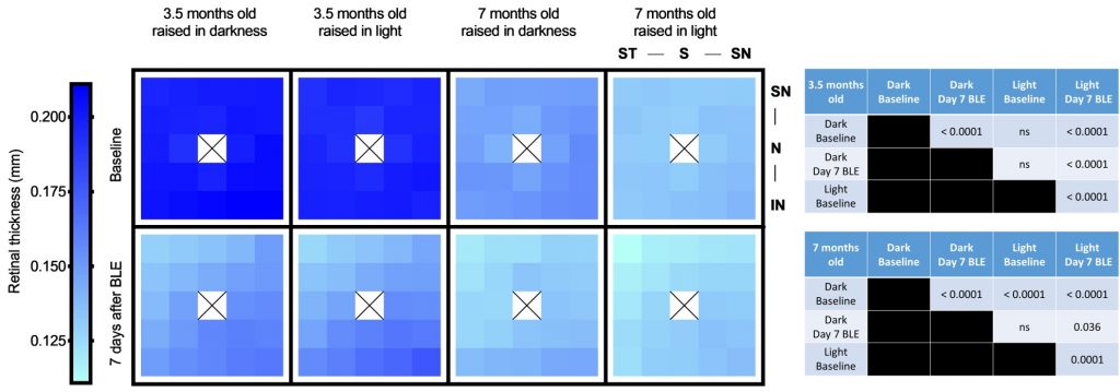
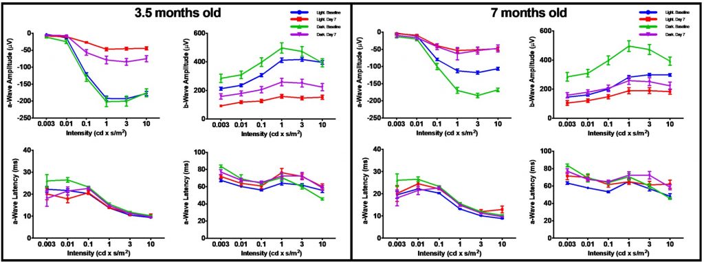
The model is induced by exposing rodents to bright light (2.500-10.000 lux). The exposure to bright light (BLE) induces the apoptosis of photoreceptors and retinal pigment epithelium1-3. Our previous studies have shown that the total retinal thickness in decreases with aging, as seen in 7 month-old vs 3.5 month-old BALB/c mice. Moreover, the BLE exacerbates the retinal damage in aged mice when compared to young mice.
| Animal species | Mice |
| Method of induction | Exposure to bright light |
| Follow-up period | Up to 7 days |
| Route of compound administration | Topical (e.g. eye drops), subretinal, intravitreal, systemic |
| Read-outs | 1. In vivo imaging: spectral-domain optical coherence tomography (SD-OCT), 2. In vivo functional assessment, 3. Morphological assessment, 4. Molecular biology (ELISA, Western blotting, qPCR) |
Read-outs
References
- Tanito M, Kaidzu S, Anderson RE. Delayed loss of cone and remaining rod photoreceptor cells due to impairment of choroidal circulation after acute light exposure in rats. Invest Ophthalmol Vis Sci 2007 vol. 48(4):1864-1872.
- Youssef PN, Sheibani N, Albert DM. Retinal light toxicity. Eye 2011 25:1-14.
- Song D, Song Y, Hadziahmetovic M, Zhong Y, Dunaief JL. Systemic administration of the iron chelator deferiprone protects against light-induced photoreceptor degeneration in the mouse retina. Free Radical Biol Med 2012 53:64-71