


Summary: OIR model mimics retinopathy of prematurity (ROP) and human ischemic retinopathies.
Model Description
Oxygen-induced retinopathy model mimics retinopathy of prematurity (ROP) and human ischemic retinopathies. Two rodent models, mouse and rat, are commonly used to study the pathophysiology of ROP and to test the preclinical efficacy of drug candidates for ROP. However, mouse and rat oxygen-induced retinopathy (OIR) models differ in their mode of induction. In mice, chronic hyperoxia is induced versus alternating hyperoxia/hypoxia in rats. The manifestation of their vascular phenotypes also is different. In mice, vaso-obliteration occurs primarily in the central retina. In contrast, vaso-obliteration is more peripheral in the rat oxygen-induced retinopathy model and in human ROP (Kim, D’Amore and Connor, 2016).
At Experimentica, we have fully implemented and validated both mouse and rat OIR models with a clinically relevant reference compound and the most comprehensive list of read-outs. More information about both mouse and rat OIR model can be shared on demand.
| Animal species | Mice, Rats |
| Method of induction | Exposure to high oxygen levels during retinal vasculature development |
| Follow-up period | Typically 5-6 days after the animals are returned into normoxic condition |
| Route of compound administration | Topical (e.g. eye drops), intravitreal injection, systemic |
| Read-outs | Morphological assessment of retinal vasculature using confocal microscopy: – Avascular area/vascular obliteration, – Area of neovascular tufts, – Microglial activation. |
Outcomes and Read-Outs
In vivo imaging
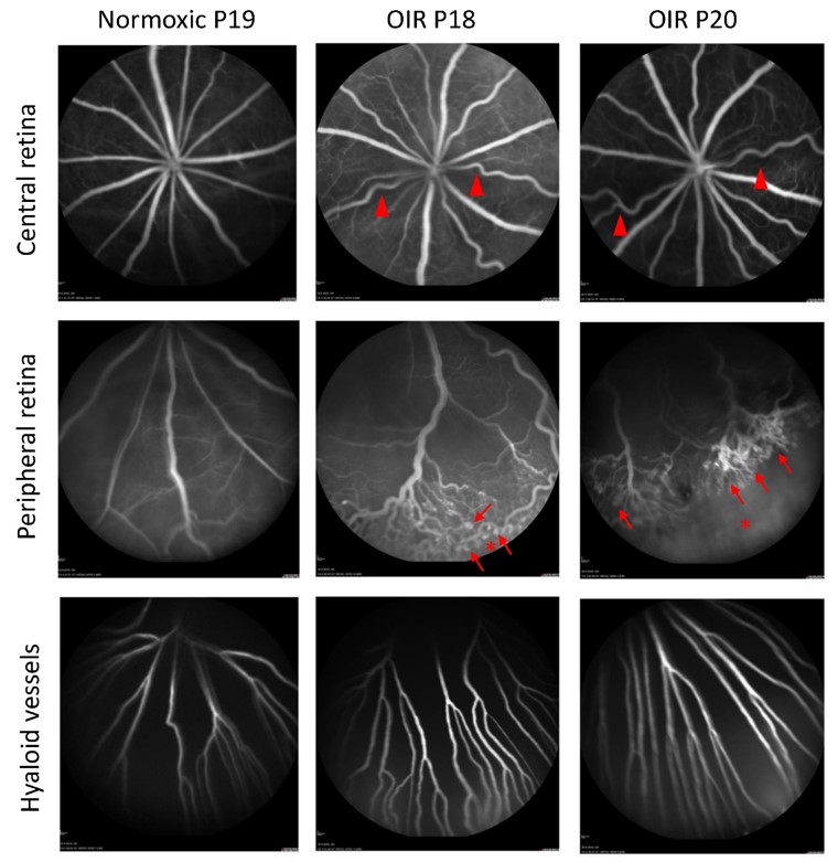
Fluorescein angiography (rat; Heideleberg Spectralis HRA2). At later timepoints of OIR development, P18-P20, pre-retinal neovascular tufts and avascular areas can be imaged in the periphery (Fig. 1).
SD-OCT (Envisu R2210 and Envisu R2210, Bioptigen Inc./Leica Microsystems) allows imaging of retinal layers and picks neovascularization in the inner retina.
Functional assessment
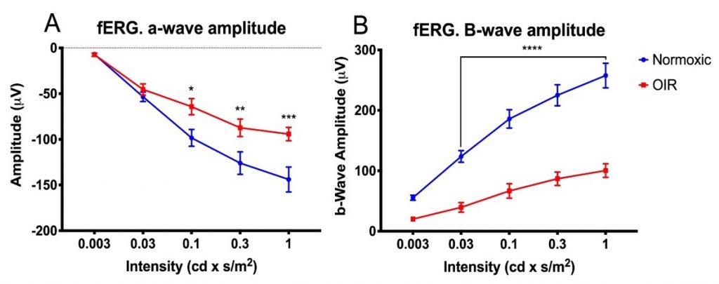
Flash electroretinography (fERG). Functional deficits are observed both in a-wave and in b-wave amplitudes in the rat OIR model at P21.
Histology/morphometry
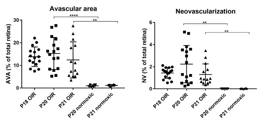
Avascular areas and areas of neovascularization are quantified from retinal wholemounts. Fig. 3 depicts the change in avascular area and neovascularization in the rat OIR model.
References
- Vähätupa M, Hakkarainen JJ, Kaja S, Uusitalo H, Järvinen TA, Uusitalo-Järvinen H, Kalesnykas G (2018) Aflibercept inhibits physiological revascularization and pathological neovascularization in the mouse and rat oxygen-induced retinopathy models. Poster presentation at EVER 2018 conference.
- Kaja S, Ragauskas S, Vähätupa M, Cerrada-Gimenez M, Mering S, Jakkarainen JJ, Kalesnykas G (2018) Standardization and validation of intravitreal and systemic administration of aflibercept in preclinical models for angiogenesis. Poster presentation at ARVO 2018 conference.
- Smith LE, et al., Oxygen-induced retinopathy in the mouse. Invest Ophthalmol Vis Sci, 1994. 35(1): p. 101-11.
- Kim CB, D’Amore PA, Connor KM. Revisiting the mouse model of oxygen-induced retinopathy. Eye Brain, 2016. 8: p. 67-79.