In vivo models
Laser-Induced Glaucoma
Two widely used ocular hypertension models for studying glaucoma pathophysiology and evaluating potential therapies are the magnetic microbead-induced elevated intraocular pressure (IOP) model and the episcleral vein photocoagulation model.
The magnetic microbead-induced model was first described by Samsel and colleagues in 2011. The elevated IOP is induced by injecting magnetic particles into the mouse or rat anterior chamber to obstruct aqueous outflow, leading to a gradual and sustained increase in intraocular pressure (IOP). The episcleral vein photocoagulation model, developed by Shareef and colleagues in 1995, induces IOP elevation through targeted coagulation of episcleral veins, creating a chronic hypertensive environment that closely mimics the gradual progression of glaucoma in humans. Both methods effectively mimic glaucomatous damage, allowing for the assessment of neuroprotective and IOP-lowering therapies. In addition, these models are particularly useful for studying chronic retinal ganglion cell (RGC) degeneration and optic nerve damage.
Recent advances in in vivo imaging and functional assessment have further enhanced both models, making them valuable and complementary tools for glaucoma research. Their ability to replicate key aspects of human disease progression enables precise evaluation of novel therapeutic strategies.
Technical details
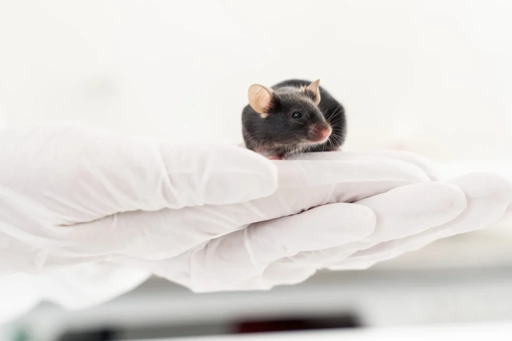
Rat
Episcleral vein photocoagulation
Typically 21-28 days
Topical, intravitreal, intracameral and subretinal injection, systemic
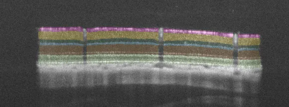
– Quantitative evaluation of inner retinal layer thickness using SD-OCT and AI-driven algorithms
– Functional RGC assessment (pERG)
– Behavioral assessment using visual acuity and contrast sensitivity (OMR)
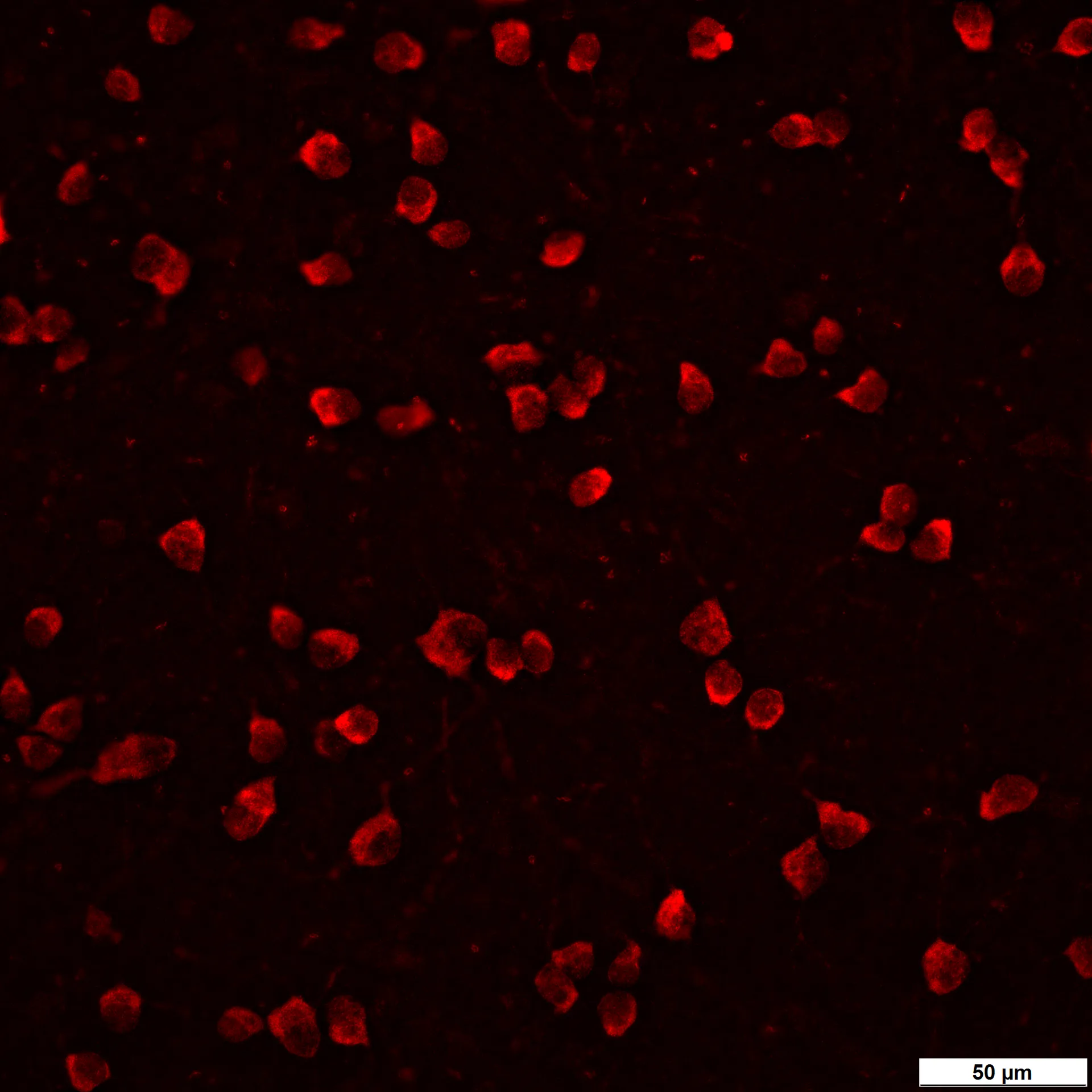
– Quantitative estimation of RGCs
Highlights of this model
Chronic IOP elevation
It is one of the best models to mimic hypertensive glaucoma.
Non-invasive monitoring of pathology
Application of 3Rs using in vivo imaging, functional assessment and behavioral analysis.
Unbiased quantitative data
From functional to morphological read-outs.
References
- Samsel, P. A., Kisiswa, L., Erichsen, J. T., Cross, S. D., & Morgan, J. E. (2011). A novel method for the induction of experimental glaucoma using magnetic microspheres. Investigative ophthalmology & visual science, 52(3), 1671-1675.
- Tribble, J. R., Otmani, A., Kokkali, E., Lardner, E., Morgan, J. E., & Williams, P. A. (2021). Retinal ganglion cell degeneration in a rat magnetic bead model of ocular hypertensive glaucoma. Translational Vision Science & Technology, 10(1), 21-21.
- Garcia-Valenzuela, E., Shareef, S., Walsh, J., & Sharma, S. C. (1995). Programmed cell death of retinal ganglion cells during experimental glaucoma. Experimental eye research, 61(1), 33-44.
Receive model details
Interested to learn more? Fill out the form below and we will email you a white paper on the disease model. Your information will not be added to any mailing lists or used for marketing purposes.
"*" indicates required fields
We are here to help
Whether you have a question about our preclinical models, capabilities, pricing or anything else, our team is ready to answer all your inquiries.
Related services
AI for image analysis
AI-driven image analysis for in vivo studies, delivering faster, unbiased results and deeper insights for your preclinical ocular programs.
Learn moreVisual Evoked Potentials
Visual evoked potential recording is used to assess visual pathway function from retina to cortex in models of glaucoma and optic neuropathies.
Learn moreOptomotor Reflex
Experimentica offers behavioral assessment and optomotor response testing to evaluate visual acuity and contrast sensitivity in rodent models.
Learn moreCheck out our latest news and activities
All News