In vivo models
Desiccating Stress + Scopolamine-Induced Dry Eye
A widely cited early murine model of Dry Eye Disease (DED) was described by Dursun et al. in 2002, introducing a reproducible approach using desiccating stress and pharmacologic inhibition to induce ocular surface damage. Building on this model, we use a validated dry eye disease model in mice to simulate both evaporative and aqueous-deficient subtypes of DED, effectively mimicking hallmark features of human dry eye disease.
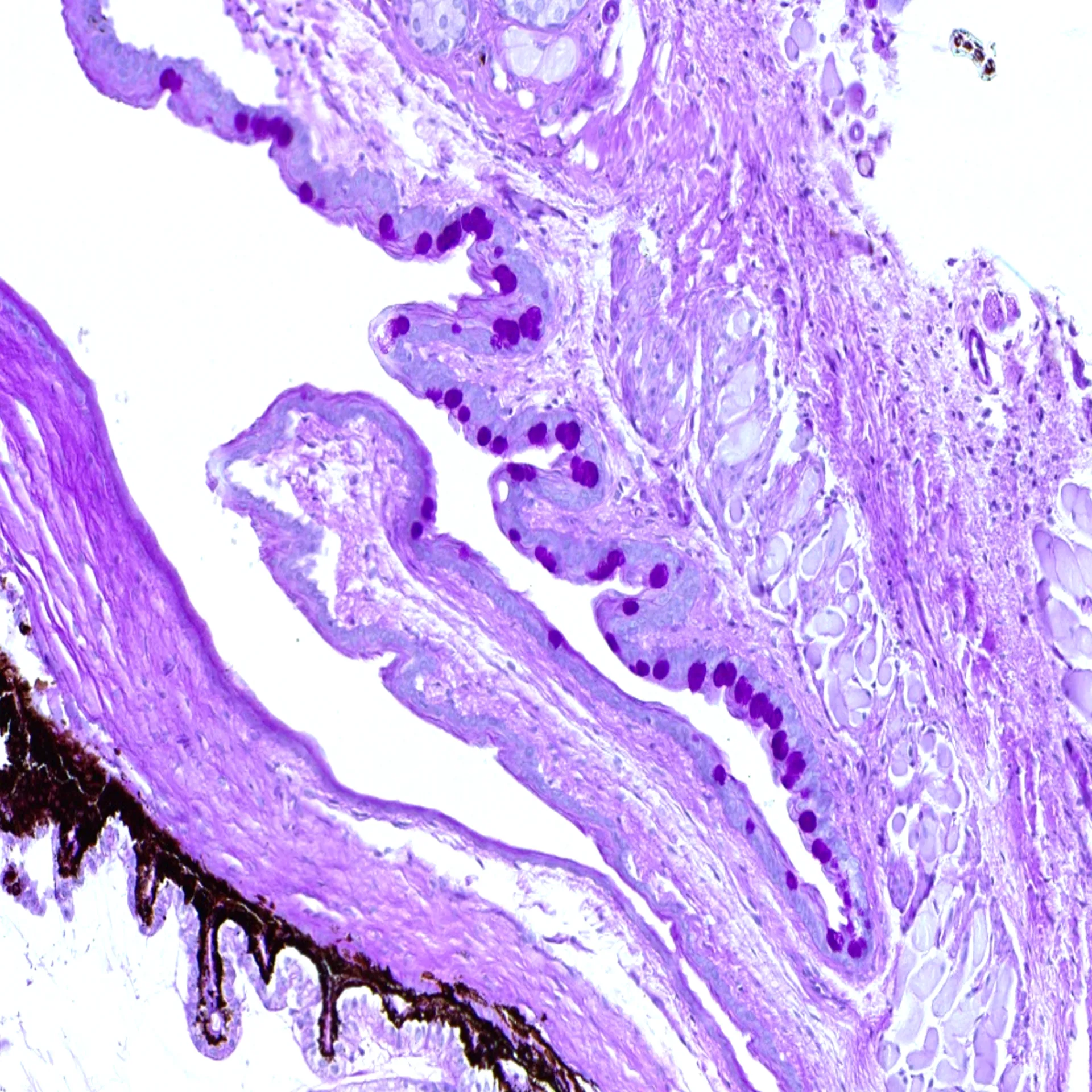
We offer comprehensive, non-invasive in vivo assessments including corneal fluorescein staining imaging, and tear volume measurements. Additionally, we provide detailed histological analysis of lacrimal gland infiltration, corneal epithelial thickness and conjunctival goblet cell density.
Technical details
Mouse
Controlled desiccating environment with scopolamine administration (injectable)
Typically, 14-21 days
In vivo assessments:
– Grading of corneal epithelial damage (corneal fluorescein staining)
– Quantitative tear volume measurement
Histological analysis:
– Grading of immune cell infiltration in the lacrimal gland
– Quantification of conjunctival goblet cells
– Quantification of corneal thickness
Highlights of this model
Scientific rigor
The model accurately mimics human DED features such as tear film instability and ocular surface damage.
Technical precision
The combination of controlled desiccating environment and pharmacologic inhibition in our model ensures reliable dry eye disease induction and consistent results throughout studies.
Comprehensive read-outs
We provide a full range of assessments, including non-invasive imaging, tear volume measurement and histology, for thorough evaluation of disease progression and treatment outcomes.
Receive model details
Interested to learn more? Fill out the form below and we will email you a white paper on the disease model. Your information will not be added to any mailing lists or used for marketing purposes.
"*" indicates required fields
We are here to help
Whether you have a question about our preclinical models, capabilities, pricing or anything else, our team is ready to answer all your inquiries.
Related services
Permeability- cornea and RPE
In vitro models using HCE-T and iPSC-RPE cells help assess drug permeability by mimicking corneal epithelium and blood-retinal barrier in early drug development.
Learn moreHistological staining
Histological staining techniques for ocular and nervous system tissues to support detailed analysis.
Learn moreImmunohistochemistry
Experimentica offers a wide range of single and multiplex immunofluorescence labeling to explore disease pathogenesis and therapeutic targets.
Learn moreRT-qPCR
qPCR measures gene expression in ocular tissues, supporting disease research and treatment evaluation.
Learn moreWestern Blotting
Western blot detects protein expression and modifications in ocular tissues with high sensitivity and precision
Learn moreELISA
ELISA enables sensitive protein detection and quantification in ocular tissues and biofluids.
Learn more