ARVO 2025 presentation: Comparison of blue light-induced damage (BLD) in albino and pigmented mice
News
05.05.2025
This study aimed at comparing blue light-induced retinal injury in albino Balb/cJRj and pigmented C57BL/6JRj mice.
News
05.05.2025
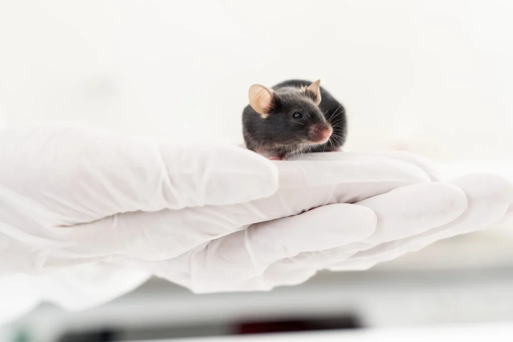
BLD was induced bilaterally to albino Balb/cJRj (5 mo old, 8 males for BLD group, 4 males for naïve group) and pigmented C57BL/6JRj mice (4 mo old, 10 males for BLD group, 4 males for naïve group). After an overnight dark adaptation and pupil dilation with tropicamide 30 min prior to induction, the mice were exposed to blue light (454 nm, 1000 lux) for 1 hour (albino) or 3 hours (pigmented).
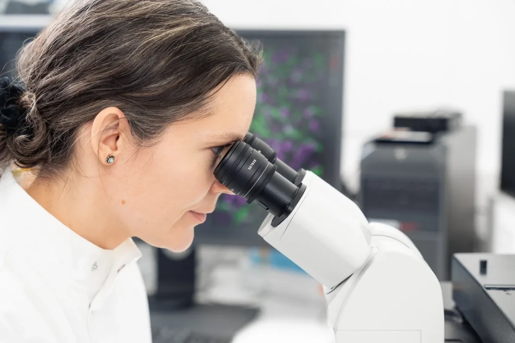
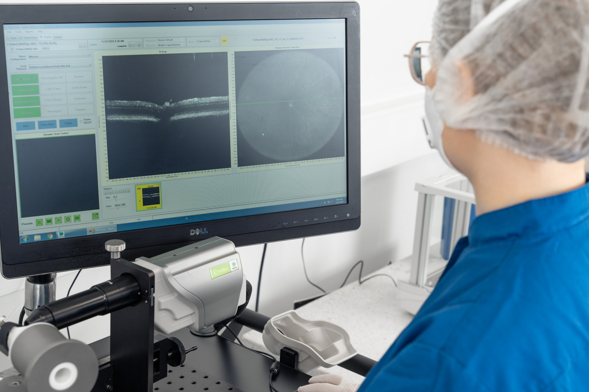
Scotopic flash electroretinography (ERG)(Celeris, Diagnosys LLC) and spectral-domain optical coherence tomography (SD-OCT)(Envisu R2200, Bioptigen Inc./Leica Microsystems) were performed at baseline and on Day 7 after induction. Retinal thickness was measured from SD-OCT images using a proprietary algorithm.
Albino mice: Seven days after BLD induction, ERG showed a significant impairment of visual function in Balb/cJRj mice. At the 0.1, 1, 3 and 10 cd.s/m2 stimuli, a-wave amplitudes were reduced between 20-43%, and b-wave amplitudes were decreased between 26-30% (t-test, P<0.01) as compared to naïve mice. The functional decline correlated with 38% retinal thinning (t-test, P<0.001) in the BLD eyes. The retinal thinning was primarily identified in the outer retina.
Pigmented mice: In C57BL/6JRj mice BLD did not affect the a- and b-wave amplitudes. Minor structural changes were seen only in the photoreceptor outer segment/retinal pigment epithelium layer (3% reduction, t-test, P<0.01).
One hour at 1,000 lux was sufficient to induce BLD in albino Balb/cJRj mice. Pigmented C57BL/6JRj mice remain resistant to BLD even at the 3-hour exposure of 1,000 lux blue light, which is in accordance with previous reports.
Leena Tähtivaara, Rubina Thapa, Päivi Partanen1 Birgitta Lappeteläinen, Hajnalka Nadai, Anne Mari Haapaniemi, Olga Vergun, Xavier Ekolle, Anna Mari Koponen, Simas Bijeikis, Kalesnykas, Giedrius, Cerrada-Gimenez, Marc
Check out our latest news and activities
All NewsAll our facilities are AAALAC accredited, reflecting our commitment to the highest standards of animal care in research.
Copyright: Experimentica Ltd. 2026