Our expertise
In vivo imaging
Advances in imaging technology have led to the adaptation of clinical imaging systems to the eyes of small laboratory animal species, notably rodents, allowing for the longitudinal high-resolution in life assessment across all species from rodents to pigs.
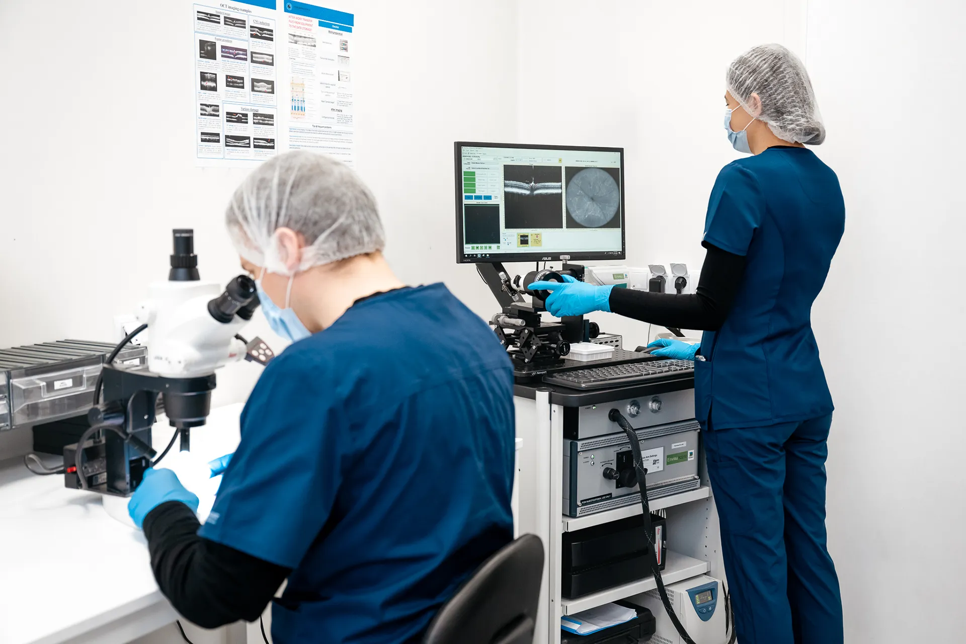
Spectral domain-optical coherence tomography (SD-OCT) is the most widely used imaging modality in clinical practice and has become the cornerstone of the in vivo assessment of the retina and the anterior segment, including the cornea and the iris, in preclinical safety and efficacy studies.
At Experimentica, we use state-of-the-art imaging equipment, including Bioptigen Envisu (Leica Microsystems) and Spectralis HRA (Heidelberg Engineering) devices to acquire SD-OCT scans. High-resolution scans can be analyzed using commercial software solutions or our proprietary algorithm to derive accurate and unbiased retinal thickness measurements.
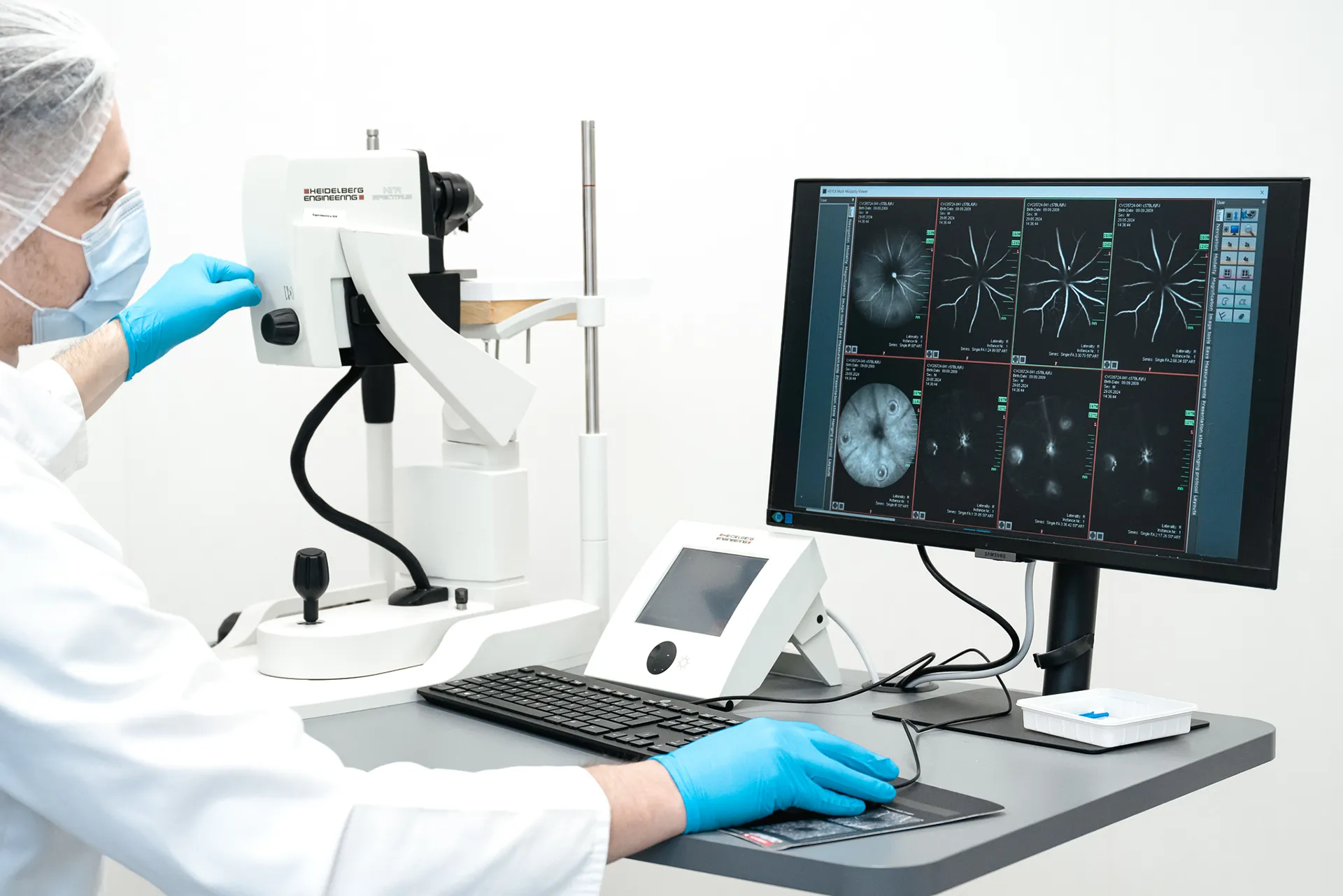
Additional imaging modalities, including fluorescein angiography (FA) and specular microscopy, can supplement the SD-OCT structural assessment, e.g. when suspecting vascular toxicity induced by test articles.
Technical details
– Corneal fluorescein staining (mouse)
– Fluorescein angiography (FA) (mouse, rat and rabbit)
– Fundus Autofluorescence (mouse and rat)
– Infrared reflectance (mouse, rat and rabbit)
– Indocyanine green angiography (ICGA) (mouse)
– Slit lamp ophthalmoscopy (mouse, rat and rabbit)
– Spectral domain optical coherence tomography (SD-OCT; mouse, rat and rabbit)
– Vitreous fluorophotometry (rat and rabbit)
We are here to help
Whether you have a question about our preclinical models, capabilities, pricing or anything else, our team is ready to answer all your inquiries.
Related services
Microbead-Induced Glaucoma
A well-established glaucoma model simulating chronic IOP elevation and enabling assessment of optic nerve damage and neuroprotective therapies.
Learn moreBlue Light Damage-Induced Retinal Degeneration
The blue light-induced retinal damage model is used for studying phototoxicity-induced retinal degeneration, and is particularly relevant for conditions such as AMD.
Learn moreLaser-Induced Choroidal Neovascularization
Laser-induced CNV model mimics exudative AMD and supports evaluation of anti-angiogenic and anti-fibrotic therapies.
Learn moreCheck out our latest news and activities
All News