Accelerate ocular safety with targeted toxicity studies
At Experimentica, we help sponsors de-risk their drug candidates early and move forward with confidence thanks to non-GLP ocular toxicity studies, available in both in vitro and in vivo models. Whether you are fine-tuning dose ranges or confirming local tolerability, our scientists deliver the data you need in a timely fashion.
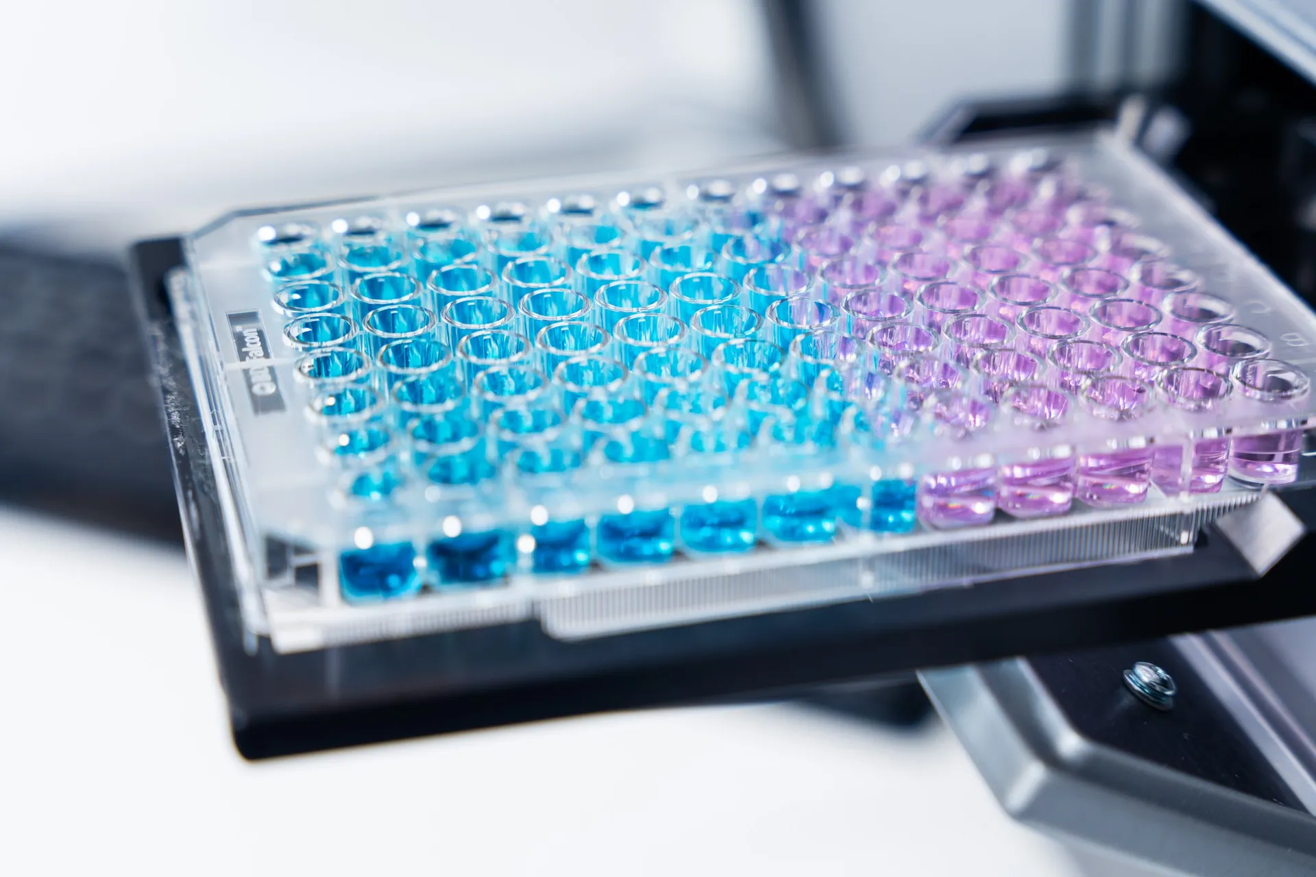
In vitro cell viability
Fast, scalable screening
Our in vitro cell viability assays are built for generating reliable safety signals before moving to animal studies. We use human corneal epithelial cells (HCE-T) and induced pluripotent stem cell-derived retinal pigment epithelium (iPSC-RPE) to test for front- and back-of-the-eye cytotoxicity. These models offer flexible exposure timelines and quantitative readouts, making them ideal for screening different drug candidates, formulations, and concentrations in early preclinical development.
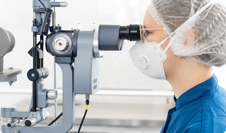
In vivo toxicity
Enabling data-driven decisions
With the right tools and methods to evaluate in vivo safety, our rodent and rabbit models combine routine clinical ophthalmic exams (slit-lamp biomicroscopy, SPOTS) with detailed structural and functional assessment of the visual system using imaging and electrophysiological modalities.
These in-life assessments are typically complemented by histopathology and biomarker quantification. Systemic and other organ-specific assessments can be added to the ocular examination.
Typical study designs include acute, sub-chronic, and chronic exposure, to explore dose escalation, MTD, and dose-range finding.
All study designs can be adjusted to meet specific requirements or help de-risk assets before entering GLP toxicity and IND-enabling studies.
Streamlined from start to finish
We have built our processes to support fast time to study start, to minimize delays and maximize clarity. Sponsors benefit from a tailored reporting that focuses on the read-outs that matter most to them so they can make decisions with confidence and keep their drug development program moving forward.
We are here to help
Whether you have a question about our preclinical models, capabilities, pricing or anything else, our team is ready to answer all your inquiries.
Related
In vivo imaging
Experimentica offers extensive in vivo imaging capabilities for high-resolution ocular assessments across species.
Learn moreFlash Electroretinography
Flash ERG is a non-invasive method to assess retinal function in preclinical eye disease models.
Learn morePattern Electroretinography
Pattern ERG is a non-invasive method to assess retinal ganglion cell function and monitor dysfunction in preclinical eye disease models.
Learn moreHistological staining
Histological staining techniques for ocular and nervous system tissues to support detailed analysis.
Learn moreImmunohistochemistry
Experimentica offers a wide range of single and multiplex immunofluorescence labeling to explore disease pathogenesis and therapeutic targets.
Learn more