In vivo models
White Light Damage-Induced Retinal Degeneration
The white light-induced retinal damage is widely used for studying retinal degeneration. Exposure to high-intensity white light induces oxidative stress, leading to photoreceptor death, retinal pigment epithelium (RPE) dysfunction, and neuroinflammation (Grimm and Remé, 2019).
This model is a valuable tool for evaluating potential therapeutic agents for retinal degeneration, oxidative stress responses, and neuroprotective strategies. It is particularly relevant for conditions such as retinitis pigmentosa (RP) and age-related macular degeneration (AMD) and other retinal diseases associated with light-induced damage (Organisciak and Vaughan, 2010). The white light model effectively mimics aspects of retinal degeneration, making it a useful system for preclinical studies on retinal protection and repair (Cerrada-Gimenez et al, 2024).
Technical details
Geographic Atrophy, Dry AMD, Retinitis Pigmentosa
Albino mouse
White LED light exposure
7 days
Systemic, topical and intravitreal
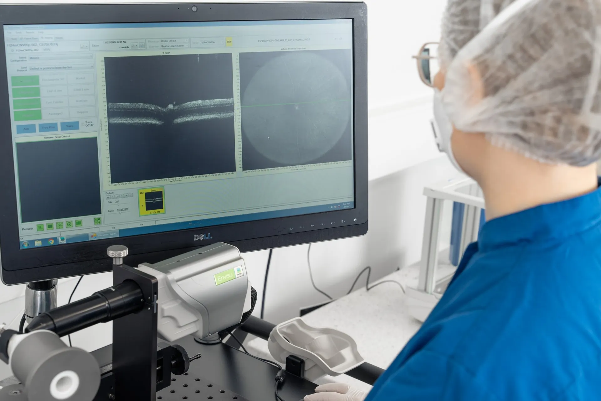
– Retinal thickness from SD-OCT scans and AI-driven analysis
– Functional assessment using ERG
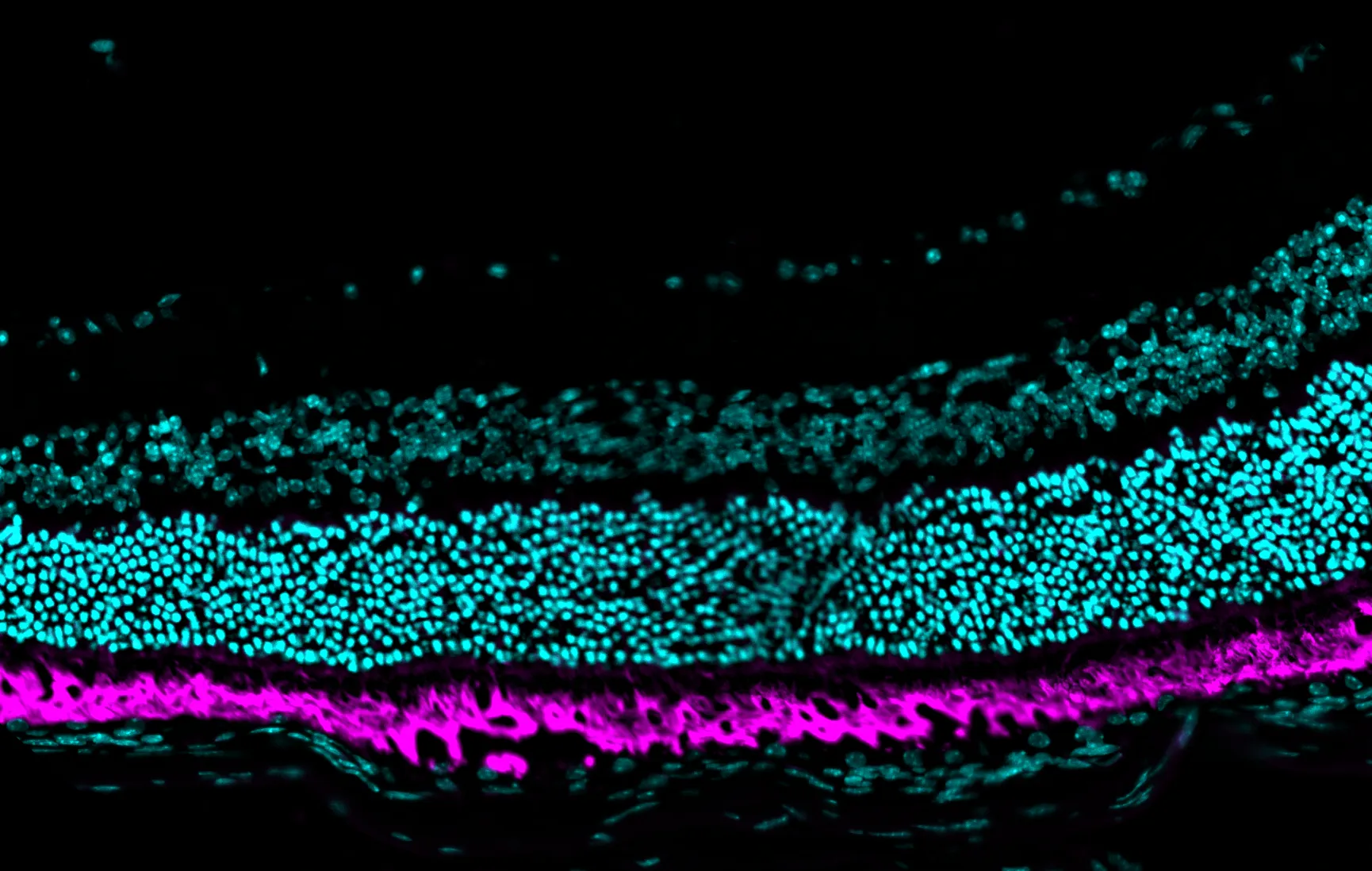
– Histology and immunohistochemistry
Highlights of this model
Rapid screening of neuroprotective properties
Quick turnaround and standardized readouts
Comprehensive efficacy evaluation
Advantage of multiple efficacy evaluation timepoints, incl. in vivo imaging and ERG
Advanced and unbiased data
Gain reliable insights through detailed analysis.
References
Receive model details
Interested to learn more? Fill out the form below and we will email you a white paper on the disease model. Your information will not be added to any mailing lists or used for marketing purposes.
"*" indicates required fields
We are here to help
Whether you have a question about our preclinical models, capabilities, pricing or anything else, our team is ready to answer all your inquiries.
Related services
Flash Electroretinography
Flash ERG is a non-invasive method to assess retinal function in preclinical eye disease models.
Learn moreIn vivo imaging
Experimentica offers extensive in vivo imaging capabilities for high-resolution ocular assessments across species.
Learn moreHistological staining
Histological staining techniques for ocular and nervous system tissues to support detailed analysis.
Learn moreImmunohistochemistry
Experimentica offers a wide range of single and multiplex immunofluorescence labeling to explore disease pathogenesis and therapeutic targets.
Learn moreRT-qPCR
qPCR measures gene expression in ocular tissues, supporting disease research and treatment evaluation.
Learn moreELISA
ELISA enables sensitive protein detection and quantification in ocular tissues and biofluids.
Learn moreWestern Blotting
Western blot detects protein expression and modifications in ocular tissues with high sensitivity and precision
Learn moreCheck out our latest news and activities
All News