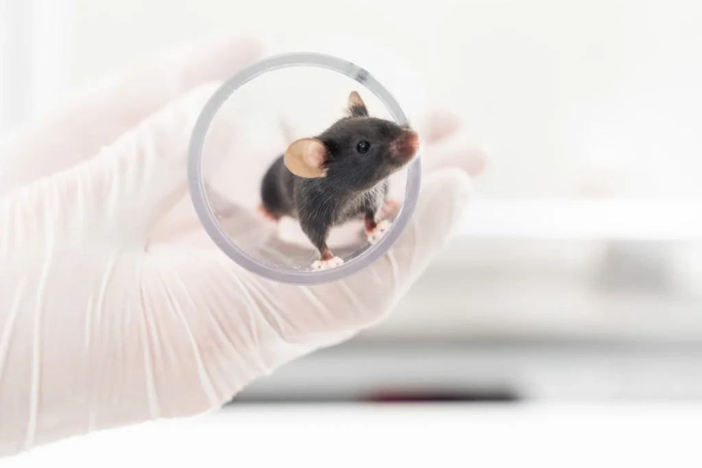
Disease models
Sodium Iodate-Induced Retinal Degeneration
The sodium iodate (NaIO3)-induced retinal degeneration model is a widely used tool for investigating age-related macular degeneration (AMD). In this model, NaIO3, administered via intravenous injection, causes oxidative stress that leads to the death of the retinal pigment epithelium (RPE) followed by secondary photoreceptor degeneration, mimicking the progression of AMD.
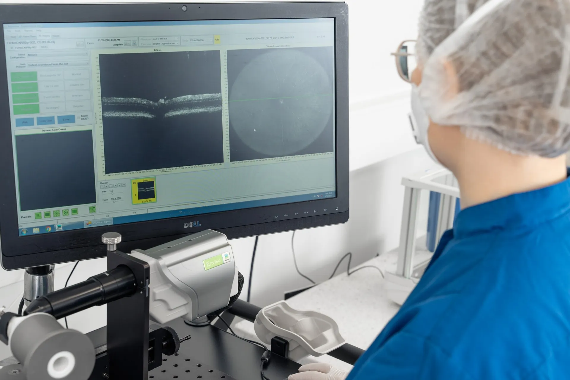
Our capabilities in this model include advanced non-invasive in vivo imaging techniques, such as fundus autofluorescence (FAF) to monitor retinal degeneration, spectral-domain optical coherence tomography (SD-OCT) for quantitative assessment of individual retinal layer thickness, and flash electroretinography (fERG) to assess retinal function, providing a comprehensive platform for evaluating potential AMD therapies.
Technical details
Rat and rabbit
IV injection of sodium iodate
Typically up to 35 days
In vivo imaging
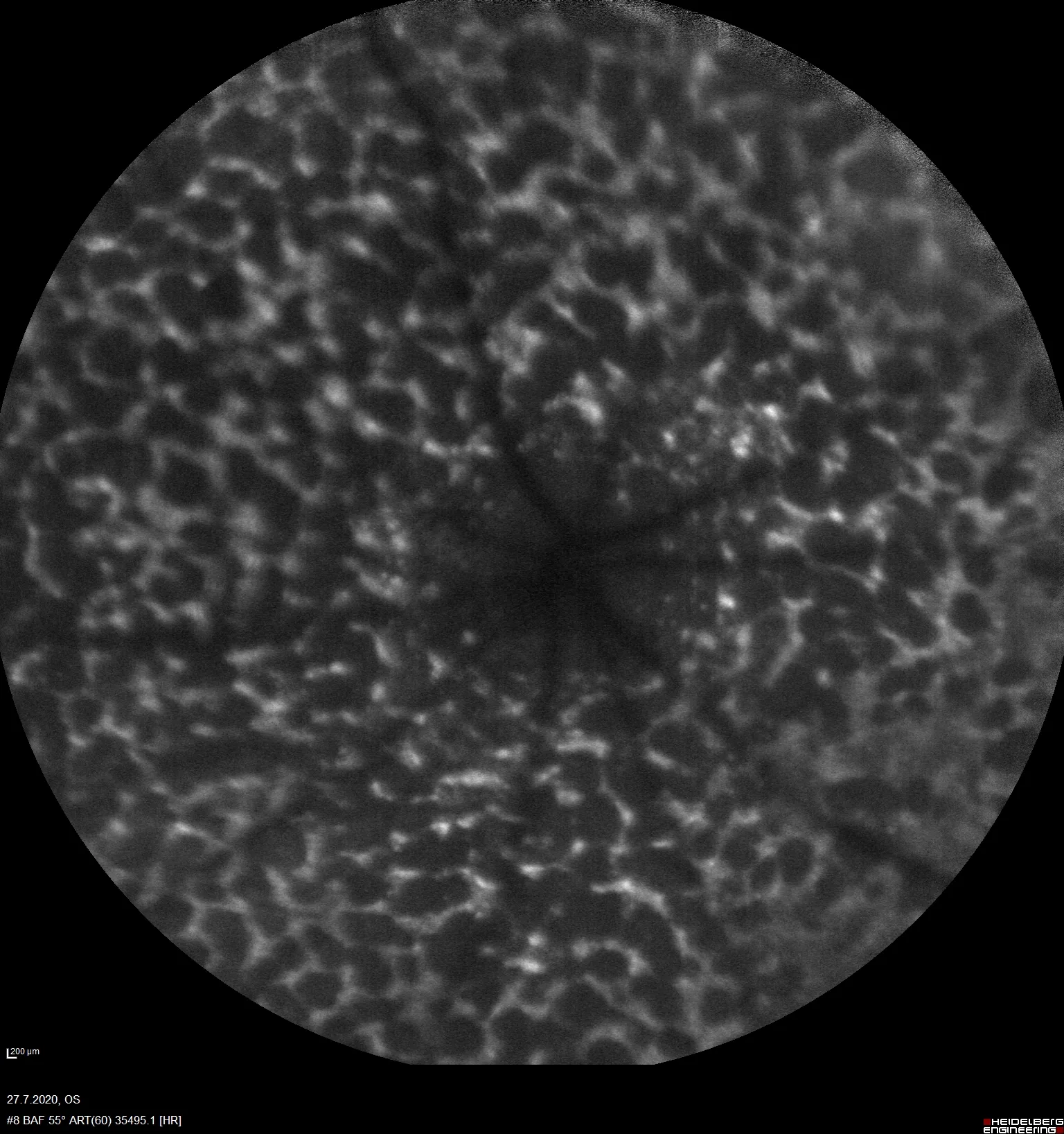
– Fundus autofluorescence for visualization of damaged area
– Spectral-domain optical coherence tomography for retinal thickness
– Electroretinography for rod bipolar cell function protection
Immunohistochemistry
– RPE65 or other markers
Scientific excellence in every model
Relevance to Human Disease
The model replicates retinal degeneration seen in conditions affecting RPE and photoreceptors (e.g. AMD).
Advanced Monitoring
Non-invasive imaging (OCT, FA) allows detailed structural and functional tracking of retinal changes.
Cost-Effective and Reliable
A well-established, efficient model for preclinical therapy testing in retinal degeneration.
Access model details
Interested to learn more? Submit the form, and we will provide tailored information on this model.
"*" indicates required fields
We are here to help
Whether you have a question about our preclinical models, capabilities, pricing or anything else, our team is ready to answer all your inquiries.
Related services
Flash Electroretinography
Flash ERG is a non-invasive method to assess retinal function in preclinical eye disease models.
Learn moreIn vivo imaging
Experimentica offers extensive in vivo imaging capabilities for high-resolution ocular assessments across species.
Learn moreHistological staining
Histological staining techniques for ocular and nervous system tissues to support detailed analysis.
Learn moreImmunohistochemistry
Experimentica offers a wide range of single and multiplex immunofluorescence labeling to explore disease pathogenesis and therapeutic targets.
Learn moreRT-qPCR
qPCR measures gene expression in ocular tissues, supporting disease research and treatment evaluation.
Learn moreWestern Blotting
Western blot detects protein expression and modifications in ocular tissues with high sensitivity and precision
Learn moreELISA
ELISA enables sensitive protein detection and quantification in ocular tissues and biofluids.
Learn moreCheck out our latest news and activities
All News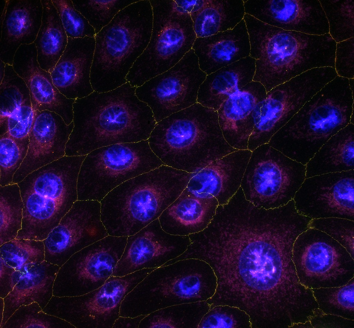
Faster results for in vitro corneal permeability studies

Experimentica Appoints Dr. Artem Shatillo as Director, Digital Transformation
FELASA 2025 presentation: Assessing lidocaine-based analgesia for mouse ear notching: Insights into strain-specific reactions