Ocular pharmacokinetics
Understanding how your drug candidate behaves in the eye is essential to its successful development. What sets Experimentica apart in ocular pharmacokinetic (PK) studies are these two complementary sets of skills: ocular administration and sub-ocular tissue collection.
A full spectrum of ocular administration routes
From small molecules to complex biologics, we support nearly every ophthalmic route of administration, including:
- Topical (eye drops)
- Intravitreal
- Subretinal
- Suprachoroidal
- Intracameral
- Subconjunctival
- Intragastric gavage (PO), IV, IP, IM
From small molecules in solutions to sustained-release implants, our team ensures accurate and reproducible dosing across mice, rats, and rabbits.
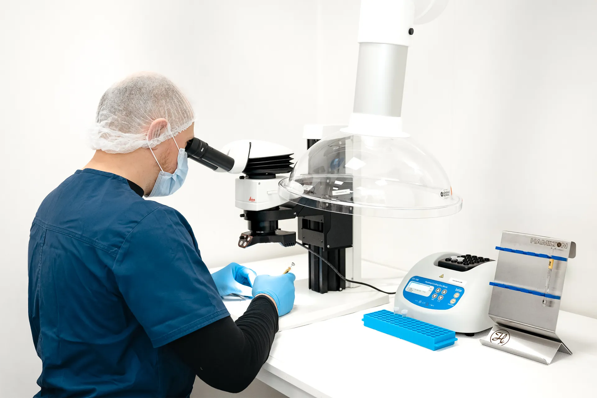
Dissection & sample collection
Our scientists are trained in advanced microdissection techniques to isolate specific ocular sub-tissues depending on species, including but not limited to cornea, lens, iris and ciliary body, aqueous humor, vitreous humor, retina, RPE+choroid, sclera, and optic nerves.
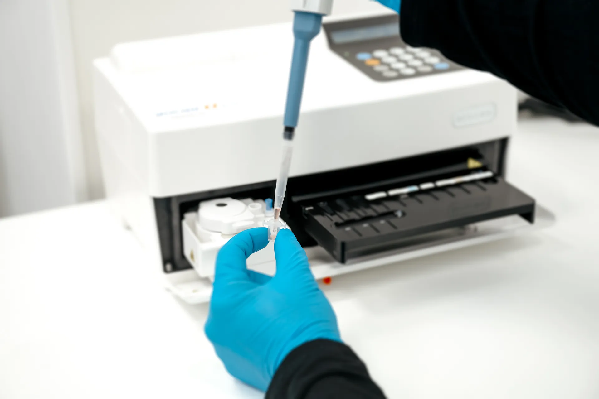
Streamlined data generation with bioanalytical partners
Our dedicated scientific team helps our sponsors move from study design to conclusion without delays. Experimentica coordinates seamlessly with our network of trusted bioanalytical partners for sample bioanalysis, so sponsors get results they can act on.
We built our PK platform for precision, reliability, and speed, with the following in mind:
- Partnering with bioanalytical experts
- Rapid in vivo study starts and short lead times
- Custom sampling schedules designed to maximize data per animal
- Flexible protocols for single or repeat dosing
We are here to help
Whether you have a question about our preclinical models, capabilities, pricing or anything else, our team is ready to answer all your inquiries.
Related
Tissue processing
Specialized processing of ocular and neural tissues employing methods such as flat mounting, paraffin and cryo embedding, and precision sectioning.
Learn more