Ocular Tolerability and Safety studies

Ocular safety studies in preclinical laboratory animal species are conducted to evaluate the tolerability and potential toxicity of test articles on the eye. Experimentica specializes exclusively in non-GLP ocular safety studies in rodents, guinea pigs, rabbits, and minipigs.

Ocular tolerability and safety can be determined by clinical evaluation, i.e. ophthalmic exams, as well as the detailed in vivo structural and functional assessment of the visual system using imaging and electrophysiological modalities. These in life assessments are typically complemented by histopathology. Systemic and other organ-specific assessments can be added to the ocular examination.

Typical study designs include the evaluation of acute, sub-acute and chronic local toxicity, determination of the maximum tolerated dose (MTD), and dose-range finding. All study designs can be adjusted to meet specific requirements or help de-risk assets before entering IND-enabling studies.

Ophthalmic Examinations

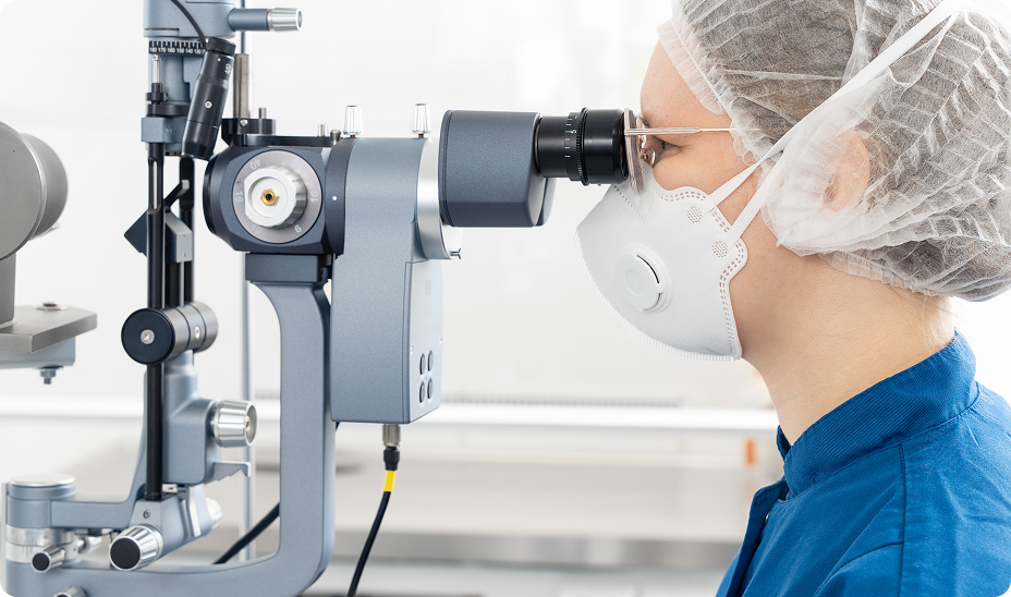
The ophthalmic examination (OE) is a standardized method for the evaluation of the entire eye that includes both a macroscopic evaluation of the outside of the eye, including the lids, orbit, and surrounding tissue and a functional assessment of ocular reflexes. This is followed by a detailed investigation of the anterior and posterior segments of the eye by slit-lamp and ophthalmoscopy.

Various protocols exist to provide a standardized scoring system of the clinical observations:

  • SPOTS method1
  • Modified McDonald-Shadduck score2
  • Modified Hackett-McDonald score3

The SPOTS method (short for “semiquantitative preclinical ocular toxicology scoring”) was developed to address the needs for standardization of preclinical drug development and toxicology programs based on modern principles and accepted standards of comparative ophthalmology. As such, SPOTS offers a more granular scoring system, which is particularly useful for the assessment of topically and intravitreally-delivered test articles.

Ocular tolerability and safety studies are performed by veterinary ophthalmologists, veterinarians and trained researchers.

Functional assessment of the visual system

Electroretinography (ERG) measures the electrical activity associated with the phototransduction cascade in the retina following a light stimulus. Different stimulation protocols using either light flash or pattern stimuli can dissociate individual components of the retinal response that can be correlated to the function of distinct retinal cell types.

In clinical practice and safety studies, flash electroretinography (fERG) is the most widely-adapted ERG modality screen for retinal dysfunction and protocols follow the guidelines established by the International Society for Clinical Electrophysiology of Vision (ISCEV) for full-field clinical electroretinography4. fERG-associated waveforms consist of an a-wave, representing the photoreceptor response, and a b-wave associated with photoreceptor/bipolar cell activity, which serve as the primary readouts for safety studies. Specialized protocols can evaluate oscillatory potentials, photopic negative response (PhNR), c-waves and d-waves, however, these parameters are typically only evaluated in disease models or to identify toxicity associated with specific cell types.

Figure 2. Representative ERG waveforms in various animal species. A: Mouse, scotopic ERG, dimmest to brightest flash intensity (200µV/100ms). B: Rat, scotopic ERG, dimmest to brightest flash intensity (200µV/100ms). C: Rabbit, scotopic ERG, brightest flash intensity (1,000 cd.s/m²).

Pattern electroretinography (pERG) is used to quantify retinal ganglion cell (RGC) function. This ERG modality is widely used in animal models for optic nerve disease associated with the loss of retinal ganglion cells, including glaucoma and non-arteritic ischemic optic neuropathy (NAION).

Visual evoked potentials (VEP) are a representation of the electrical activity recorded in the visual cortex after light flash or pattern stimulation. VEPs allow detection of dysfunction of or damage to the visual pathways including the optic nerve. Typically, VEP recordings are made in efficacy models involving optic nerve conduction deficits, such as optic neuropathy and optic neuritis.

Structural assessment of the visual system

The structural assessment complements the functional assessment of the eye and visual system. While the histopathological analysis of post-mortem tissues remains a key deliverable for safety studies, advances in imaging technology have led to the adaptation of clinical imaging systems to the eyes of small laboratory animal species, notably rodents, allowing for the longitudinal high-resolution in life assessment across all species from rodents to pigs.

Spectral domain-optical coherence tomography (SD-OCT) is the most widely used imaging modality in clinical practice and has become the cornerstone of the in vivo assessment of the retina and the anterior segment, including the cornea and the iris, in preclinical safety and efficacy studies.

Examples of SD-OCT images. A: SD-OCT Dutch Belted (DB) Rabbit retina. B: C57/BL6 Mouse retina. C: Long Evans Rat retina. D: Sprague Dawley Rat retina. E: DB Rabbit cornea. F: DB Rabbit cornea and iris.
A, C, E: Spectralis HRA (Heidelberg Engineering)
B, D: Bioptigen Envisu (Leica Microsystems)
E: Visante OCT (Carl Zeiss)
Figure 3. Examples of SD-OCT images in various animal species. A: DB rabbit retina. B: C57/BL6 mouse retina. C: Long Evans rat retina. D: Sprague Dawley rat retina. E: DB rabbit cornea. F: DB rabbit cornea and iris.
A, C, E: Spectralis HRA (Heidelberg Eng.). B, D: Bioptigen Envisu (Leica). E: Visante OCT (Zeiss)
DB rabbit: Dutch Belted rabbit

At Experimentica, we use state-of-the-art imaging equipment, including Bioptigen Envisu (Leica Microsystems) and Spectralis HRA (Heidelberg Engineering) devices to acquire SD-OCT scans. High-resolution scans can be analyzed using commercial software solutions or our proprietary algorithm to derive accurate and unbiased retinal thickness measurements. Our method of quantification relies on a convolutional neural network based on U-net architecture and a transfer learning approach, which generates segmentation masks representing the individual retinal layers.

Additional imaging modalities, including fluorescein angiography (FA) and specular microscopy, can supplement the SD-OCT structural assessment, e.g. when suspecting vascular toxicity induced by test articles.

Histopathological assessment and biomarker quantification

Histopathological analysis is performed on tissues collected post-mortem. Experimentica offers a complete in-house histopathology pipeline, including acquisition of photomicrographs and quantification.

Our Cell and Molecular Pharmacology Laboratory specializes in advanced methods of tissue analysis (see also Ocular sub-tissue collection), including biomarker quantification by molecular biological (quantitative polymerase chain reaction, qPCR) and immunobiological techniques (immunohistochemistry, Western blot, ELISA, bead-based multiplex assays). Our Microscopy Division can support advanced high-resolution microcopy and electron microscopy.

References

  1. Eaton, J.S., P.E. Miller, E. Bentley, S.M. Thomasy, and C.J. Murphy, The SPOTS System: An Ocular Scoring System Optimized for Use in Modern Preclinical Drug Development and Toxicology. J Ocul Pharmacol Ther, 2017. 33(10): 718-734.
  2. McDonald, T.O. and J.A. Shadduck, Eye irritation, in Advances in Modern Toxicology, F.N. Marzulli and H.I. Maibach, Editors. 1977, Hemisphere Publishing Corp.: Washington, D.C. p. 139 – 191.
  3. Hackett, R.B. and T.O. McDonald, Assessing ocular irritation, in Dermatotoxicology, F.N. Marzulli and H.I. Maibach, Editors. 1996, Hemisphere Publishing Corp.: Washington, D.C. p. 557 – 567.
  4. Robson, A.G., L.J. Frishman, J. Grigg, R. Hamilton, B.G. Jeffrey, M. Kondo, S. Li, and D.L. McCulloch, ISCEV Standard for full-field clinical electroretinography (2022 update). Doc Ophthalmol, 2022. 144(3): 165-177. PMC9192408.