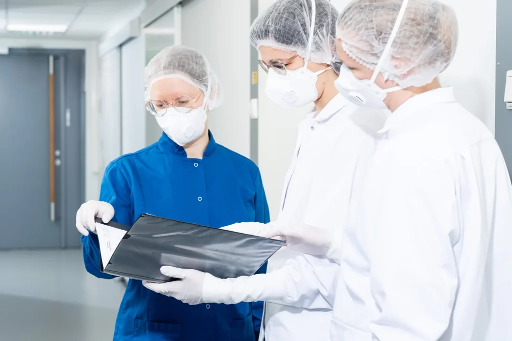
Our expertise
AI for image analysis
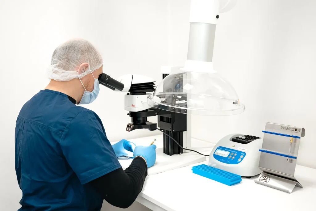
Experimentica’s expertise in AI-driven image analysis is rooted in the team’s experience with high-end imaging techniques applied to in vivo and histology readouts, along with custom algorithm and AI development.
Our internally-developed algorithms were trained on proprietary datasets and have been available and applied to sponsored studies since 2021. Experimentica’s scientists leverage AI to generate innovative read-outs and uncover new ways of interpretating existing readouts and data in a way that would otherwise be impossible to achieve manually. Our algorithms also lead to unbiased interpretation and faster generation of results and reports. Their expert oversight and quality control ensure that the generated results are both consistent and reliable.
Our mission is to provide sponsors with better results faster, which in turn enables quicker interpretation and confident decision-making for our customers.
Technical details
– Total retina thickness (mouse and rat)
– Retinal 6-layer segmentation algorithm with individual layer thickness (mouse and rat)
– Retinal 7-layer segmentation algorithm with individual layer thickness (mouse and rat)
– Laser-induced choroidal neovascularization (CNV) lesion volume analysis (mouse and rat)
– Vitreal inflammatory cell count (mouse and rat)
– Inner-outer retina thickness (mouse and rat)
– Laser-induced geographic atrophy lesion analysis (mouse)
– Sodium iodate-induced lesion analysis (mouse and rat)
– DL-AAA qualitative lesion leakage (rabbit)
– Laser-induced choroidal neovascularization (CNV) qualitative and quantitative grading of lesion leakage (mouse and rat)
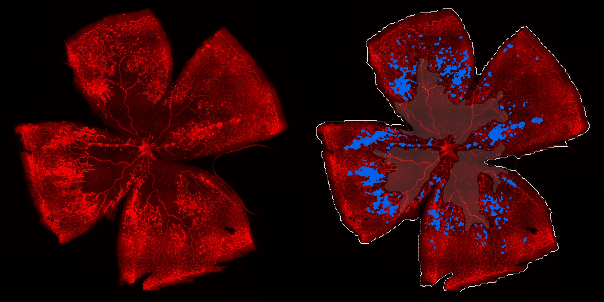
– Oxygen induced retinopathy. Quantitative evaluation of neovascular and avascular areas from Isolectin-stained retinal wholemounts (mouse)
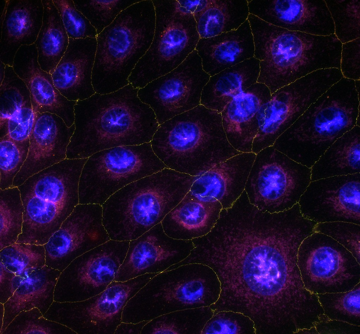
– Quantification of cells from retinal wholemounts: activated vs silent microglia, number of stained cells per area
We are here to help
Whether you have a question about our preclinical models, capabilities, pricing or anything else, our team is ready to answer all your inquiries.
Related services
Microbead-Induced Glaucoma
A well-established glaucoma model simulating chronic IOP elevation and enabling assessment of optic nerve damage and neuroprotective therapies.
Learn moreBlue Light Damage-Induced Retinal Degeneration
The blue light-induced retinal damage model is used for studying phototoxicity-induced retinal degeneration, and is particularly relevant for conditions such as AMD.
Learn moreLaser-Induced Choroidal Neovascularization
Laser-induced CNV model mimics exudative AMD and supports evaluation of anti-angiogenic and anti-fibrotic therapies.
Learn moreHistological staining
Histological staining techniques for ocular and nervous system tissues to support detailed analysis.
Learn moreImmunohistochemistry
Experimentica offers a wide range of single and multiplex immunofluorescence labeling to explore disease pathogenesis and therapeutic targets.
Learn moreCheck out our latest news and activities
All News