in vivo models
Blue Light Damage-Induced Retinal Degeneration
The blue light-induced retinal damage model is widely used for studying phototoxicity and retinal degeneration. Exposure to high-intensity blue light induces oxidative stress, leading to photoreceptor apoptosis, retinal pigment epithelium (RPE) dysfunction, and neuroinflammation (Christian and Remé, 2019).
This model is a valuable tool for evaluating potential therapeutic agents for retinal degeneration, oxidative stress responses, and neuroprotective strategies. It is particularly relevant for conditions such as age-related macular degeneration (AMD) and other retinal diseases associated with light-induced damage (Carozza et al 2024, and Chakravarthy 2024). The blue light model effectively mimics aspects of retinal pathology, making it a useful system for preclinical studies on retinal protection and repair.
Technical details
Mouse and rat
Blue LED light exposure
7 days
– Retinal thickness from SD-OCT scans
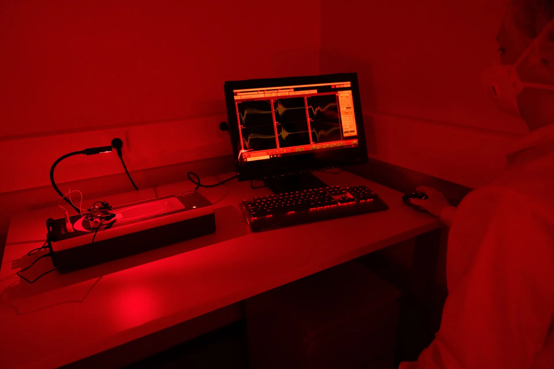
– Retina functionality from ERG
– Histology and immunohistochemistry
Multiplex labeling of the degenerating retina
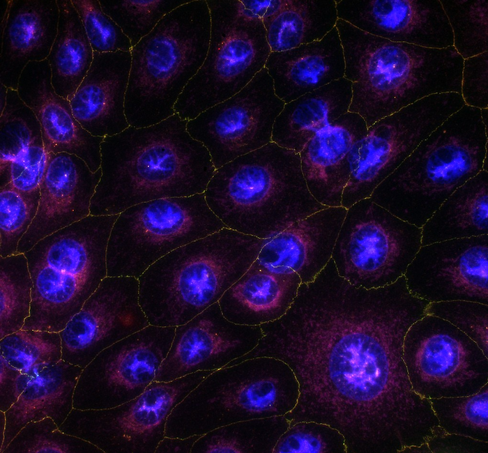
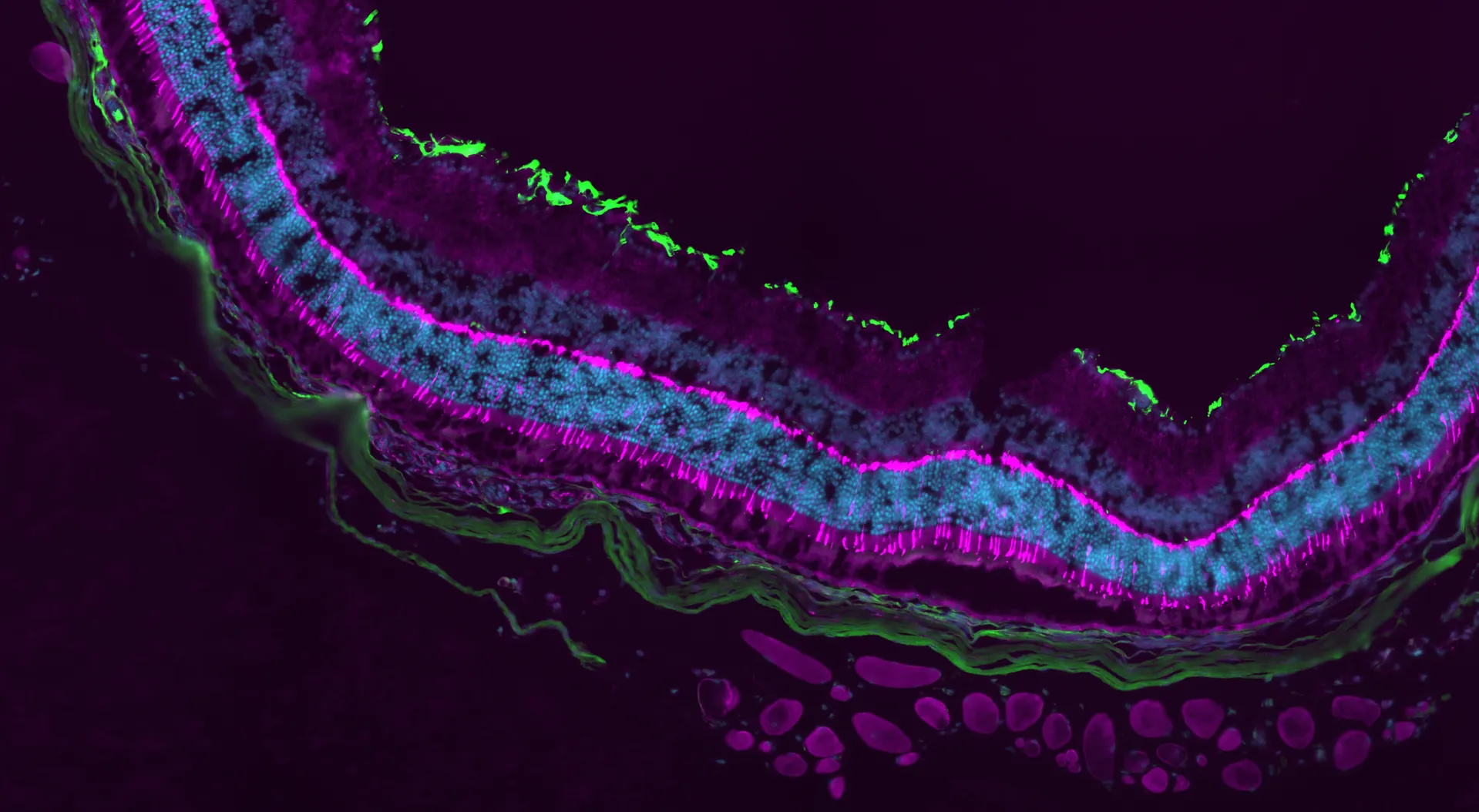
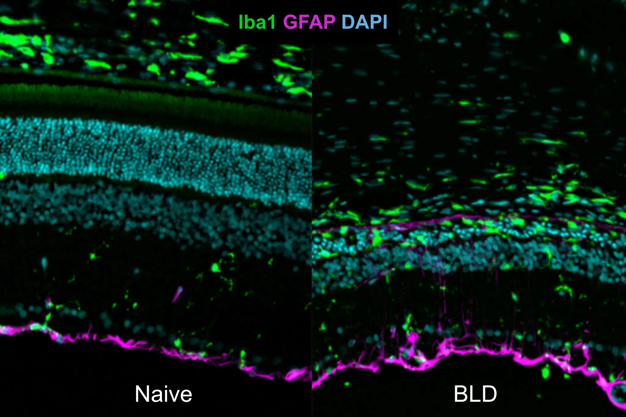
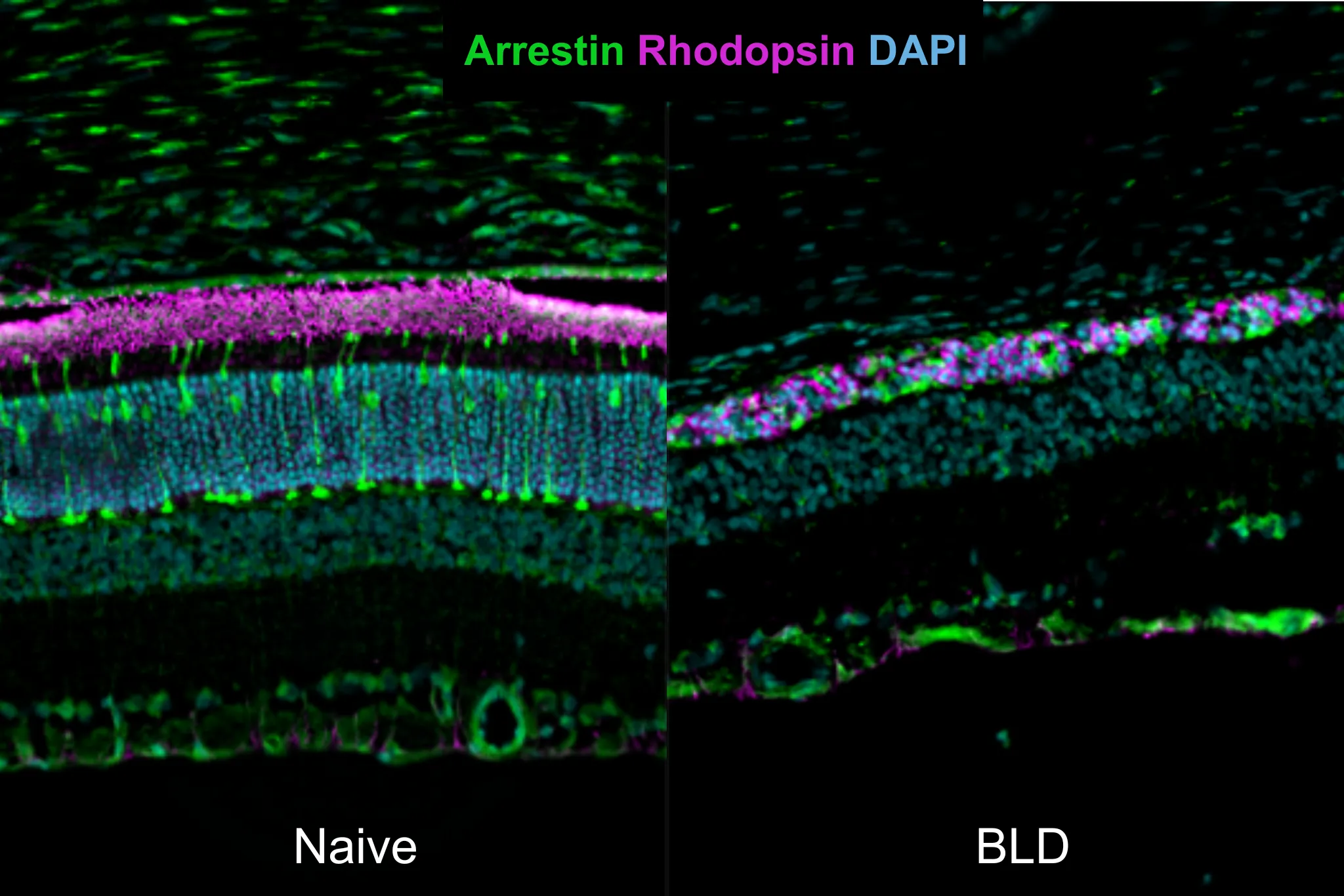
To characterize the pathological changes in the blue light damage (BLD) model for dry form of age-related macular degeneration, we offer multiplex immunofluorescence staining of eye sections. Photoreceptor integrity is evaluated using arrestin (in green in image 2) and rhodopsin (magenta). Arrestin labeling provides a robust overview of cone-specific morphology, whereas rhodopsin staining shows structural maintenance of rod outer segments. The inflammatory landscape can be visualized using Iba1 (ionized calcium-binding adapter molecule 1; green in image 1), which identifies silent and activated microglia. Additionally, glial fibrillary acidic protein (GFAP) can be utilized to monitor reactive gliosis, the upregulation of intermediate filaments in Müller cells and retinal astrocytes in response to light-induced oxidative stress.
This combinatorial labeling approach allows for the precise spatial correlation between the loss of photoreceptors and the recruitment of inflammatory mediators within the degenerative retina.
Highlights of this model
Rapid screening of neuroprotective properties
Assess the potential of your test article efficiently using our well-established in vivo model.
Comprehensive efficacy evaluation
Benefit from multiple in vivo imaging and functional analysis and timepoints.
Advanced, unbiased data analysis
Gain reliable insights through our proprietary AI-driven algorithms.
References
- Carozza, Giulia, Darin Zerti, Annamaria Tisi, Marco Ciancaglini, Mauro Maccarrone, and Rita Maccarone. 2024. “An Overview of Retinal Light Damage Models for Preclinical Studies on Age-Related Macular Degeneration: Identifying Molecular Hallmarks and Therapeutic Targets.” Reviews in the Neurosciences 35 (3): 303–30.
- Chakravarthy, Harshini, Vasil Georgyev, Cole Wagen, Amir Hosseini, and Joanne Matsubara. 2024. “Blue Light-Induced Phototoxicity in Retinal Cells: Implications in Age-Related Macular Degeneration.” Frontiers in Aging Neuroscience 16 (December).
- Grimm, Christian, and Charlotte E. Remé. 2019. “Light Damage Models of Retinal Degeneration.” Methods in Molecular Biology (Clifton, N.J.) 1834:167–78.
Receive model details
Interested to learn more? Fill out the form below and we will email you a white paper on the disease model. Your information will not be added to any mailing lists or used for marketing purposes.
"*" indicates required fields
We are here to help
Whether you have a question about our preclinical models, capabilities, pricing or anything else, our team is ready to answer all your inquiries.
Related services
Flash Electroretinography
Flash ERG is a non-invasive method to assess retinal function in preclinical eye disease models.
Learn moreIn vivo imaging
Experimentica offers extensive in vivo imaging capabilities for high-resolution ocular assessments across species.
Learn moreHistological staining
Histological staining techniques for ocular and nervous system tissues to support detailed analysis.
Learn moreImmunohistochemistry
Experimentica offers a wide range of single and multiplex immunofluorescence labeling to explore disease pathogenesis and therapeutic targets.
Learn moreRT-qPCR
qPCR measures gene expression in ocular tissues, supporting disease research and treatment evaluation.
Learn moreWestern Blotting
Western blot detects protein expression and modifications in ocular tissues with high sensitivity and precision
Learn moreELISA
ELISA enables sensitive protein detection and quantification in ocular tissues and biofluids.
Learn moreCheck out our latest news and activities
All News