in vivo models
Experimental Autoimmune Encephalomyelitis
Experimental Autoimmune Encephalomyelitis (EAE) is the most commonly used model for the preclinical development of drug candidates for treatment of multiple sclerosis1 (MS) and associated optic neuritis2.
The model is characterized by pathogenesis of autoimmunity, CNS inflammation, and demyelination. These pathologies lead to symptoms of paralysis and visual impairment. Thus, the EAE model mimics the phenotype reminiscent of MS with a well-defined onset of development, as well as aspects of the optic neuritis.
Technical details
Mouse
Injections of
– Emulsion containing MOG35-55 peptide and inactivated mycobacterium tuberculosis in complete Freuds adjuvant (CFA)
– Pertussis toxin (PTX)
28 – 42 Days
Systemic
– Clinical behavioural scoring of symptoms
– Retinal thickness assessment with SD-OCT imaging
– Assessment of visual function by electroretinogram, oscillatory potentials, photopic negative response, and/or visually evoked potentials
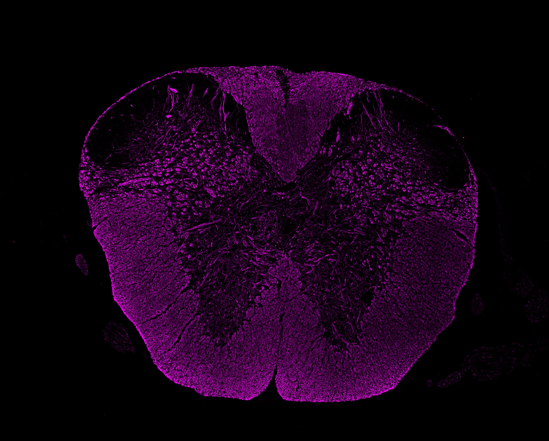
– Infiltration of optic nerve and
– Assessment of infiltration and demyelination
– Quantification of retinal ganglion cells
Highlights of this model
Preclinical MS platform
A well-established model for testing multiple sclerosis and associated optic neuritis candidates
Structure and function evaluation
Non-invasive assessment of retinal structure and visual function using advanced in vivo technologies
Neuroprotection profiling
Detailed analysis of optic nerve cellular infiltration, demyelination, and RGC loss to evaluate neuroprotective effects
References
- Huntemann N, Vogelsang A, Groeneweg L, Willison A, Herrmann A M, Meuth S G, Eichler S (2022). An optimized and validated protocol for inducing chronic experimental autoimmune encephalomyelitis in C57BL/6J mice. Journal of Neuroscience Methods. 367
- Manogaran P, Walker-Egger C, Samardzija M, Waschkies C, Grimm C, Rudin M, Schippling S. Exploring experimental autoimmune optic neuritis using multimodal imaging. NeuroImage 175, 327-339
We are here to help
Whether you have a question about our preclinical models, capabilities, pricing or anything else, our team is ready to answer all your inquiries.
Related services
Animal welfare
Learn moreVisual Evoked Potentials
Visual evoked potential recording is used to assess visual pathway function from retina to cortex in models of glaucoma and optic neuropathies.
Learn moreOptomotor Reflex
Experimentica offers behavioral assessment and optomotor response testing to evaluate visual acuity and contrast sensitivity in rodent models.
Learn moreHistological staining
Histological staining techniques for ocular and nervous system tissues to support detailed analysis.
Learn moreImmunohistochemistry
Experimentica offers a wide range of single and multiplex immunofluorescence labeling to explore disease pathogenesis and therapeutic targets.
Learn moreELISA
ELISA enables sensitive protein detection and quantification in ocular tissues and biofluids.
Learn moreRT-qPCR
qPCR measures gene expression in ocular tissues, supporting disease research and treatment evaluation.
Learn moreWestern Blotting
Western blot detects protein expression and modifications in ocular tissues with high sensitivity and precision
Learn moreCheck out our latest news and activities
All News