In vivo models
High IOP-Induced Ischemia-Reperfusion Injury
In 1996, Adachi et al. introduced a rat model of high intraocular pressure-induced ischemia and reperfusion injury induced by acutely increasing the intraocular pressure for a period of time. Accute increase of the intraocular pressure mimics primary open-angle glaucoma & acute angle-closure glaucoma by causing formation of reactive oxygen species, breakdown of inner blood-retinal barrier, and eventually retinal ganglion cell death.
Recent advancements in in vivo imaging and electrophysiology turned this model into a powerful and validated tool for the screening of novel drug candidates with neuroprotective properties. Further innovations in AI-driven image recognition offers more precise and accurate screening of novel drug candidates.
Technical details
Rat
Rapid intraocular pressure increase
Typically 7-14 days
Retinal structure and function evaluation through:
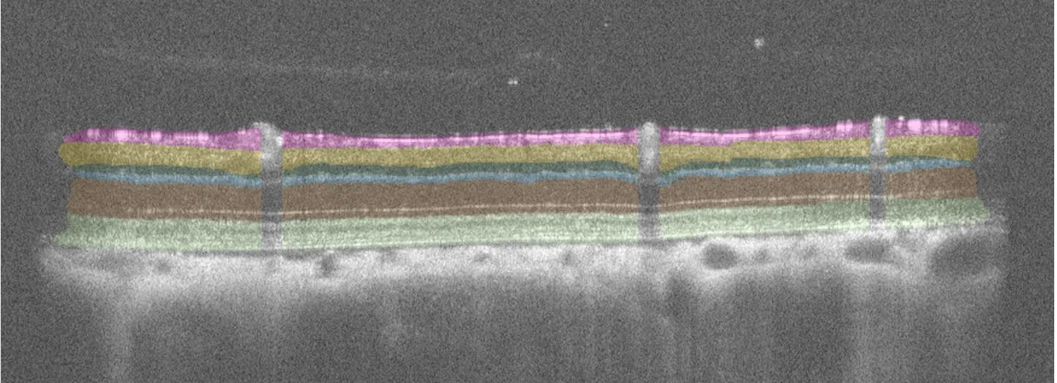
– Inner retinal thickness measurements (SD-OCT)
– Flash and pattern electroretinogram (fERG & pERG)
– Optomotor response (OMR) testing for visual acuity and contrast sensitivity
– Quantitative and unbiased estimation of RGCs from retinal flat mounts
Highlights of this model
Clinically relevant and fast
Models retinal ischemia with measurable neuronal loss within 7–14 days, enabling rapid therapeutic screening.
Precise and versatile
Controlled IOP elevation allows adjustable severity of injury, analyzed for functional and structural insights using SD-OCT, ERG, OMR and histology.
Unbiased assessment
AI-driven image analysis and stereology provide objective, reproducible RGC quantification for reliable drug efficacy assessment.
Receive model details
Interested to learn more? Fill out the form below and we will email you a white paper on the disease model. Your information will not be added to any mailing lists or used for marketing purposes.
"*" indicates required fields
We are here to help
Whether you have a question about our preclinical models, capabilities, pricing or anything else, our team is ready to answer all your inquiries.
Related services
Optomotor Reflex
Experimentica offers behavioral assessment and optomotor response testing to evaluate visual acuity and contrast sensitivity in rodent models.
Learn moreAI for image analysis
AI-driven image analysis for in vivo studies, delivering faster, unbiased results and deeper insights for your preclinical ocular programs.
Learn morePattern Electroretinography
Pattern ERG is a non-invasive method to assess retinal ganglion cell function and monitor dysfunction in preclinical eye disease models.
Learn moreCheck out our latest news and activities
All News