In vivo models
Retinitis Pigmentosa
Retinitis Pigmentosa (RP) is a group of inherited diseases that result in retinal dysfunction and gradual photoreceptor loss. Mice with mutations known to cause the disease in human patients provide a valuable model to study and test gene therapy candidates striving to maintain visual function.
At Experimentica we offer studies with Pde6brd1 and RhoP23H (B6.129S6(Cg)-Rhotm1.1Kpal/J) mouse models. Pde6brd1 mice homozygous for the rd1 mutation show an early onset of severe retinal degeneration due to a murine viral insert and a second nonsense mutation in exon 7 of the Pde6b gene (Bowes et al.). P23H mice (The Jackson Laboratory Strain #:017628) carry the P23H mutation in the rod opsin gene (proline to histidine at codon 23), one of the most common causes of autosomal dominant retinitis pigmentosa (adRP). Heterozygous mice mimic the progressive retinal degeneration seen in patients, with early shortening of rod outer segments and photoreceptor loss by postnatal day 63. Homozygous mice display a more severe phenotype, including rapid degeneration and near-complete loss of photoreceptors by day 63 (Sakami et al.).
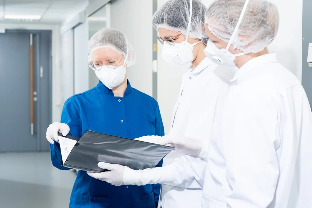
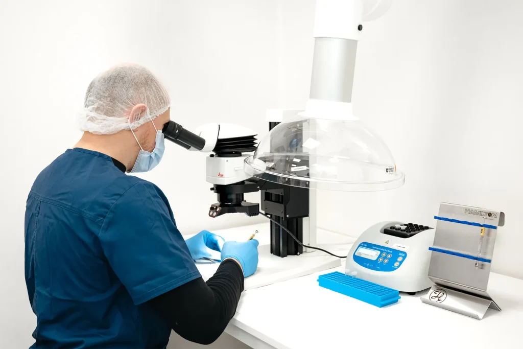
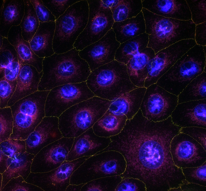
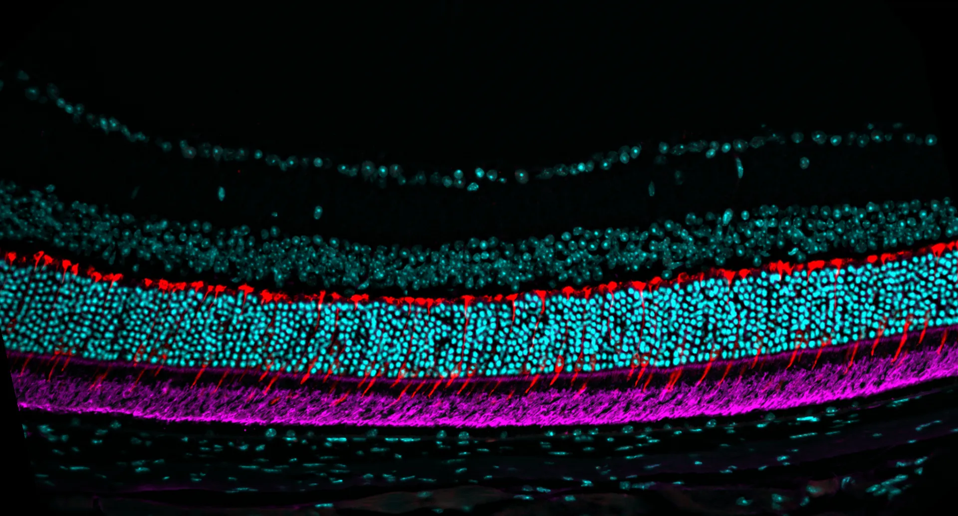
Retinal morphology is monitored utilizing spectral-domain optical coherence tomography (SD-OCT), while visual function is evaluated using flash electroretinography (fERG) and optomotor response (OMR). These non-invasive techniques enable comprehensive monitoring of disease progression with longer follow-up periods. Additionally, we provide histology and immunohistochemical stainings, enabling detailed analysis of tissue morphology and molecular markers to assess disease progression and treatment efficacy.
Technical details
Mouse
Disease onset as early as P14 (Pde6brd1), follow-up up to 12 months
Subretinal, intravitreal, systemic
– Retinal thickness (SD-OCT)
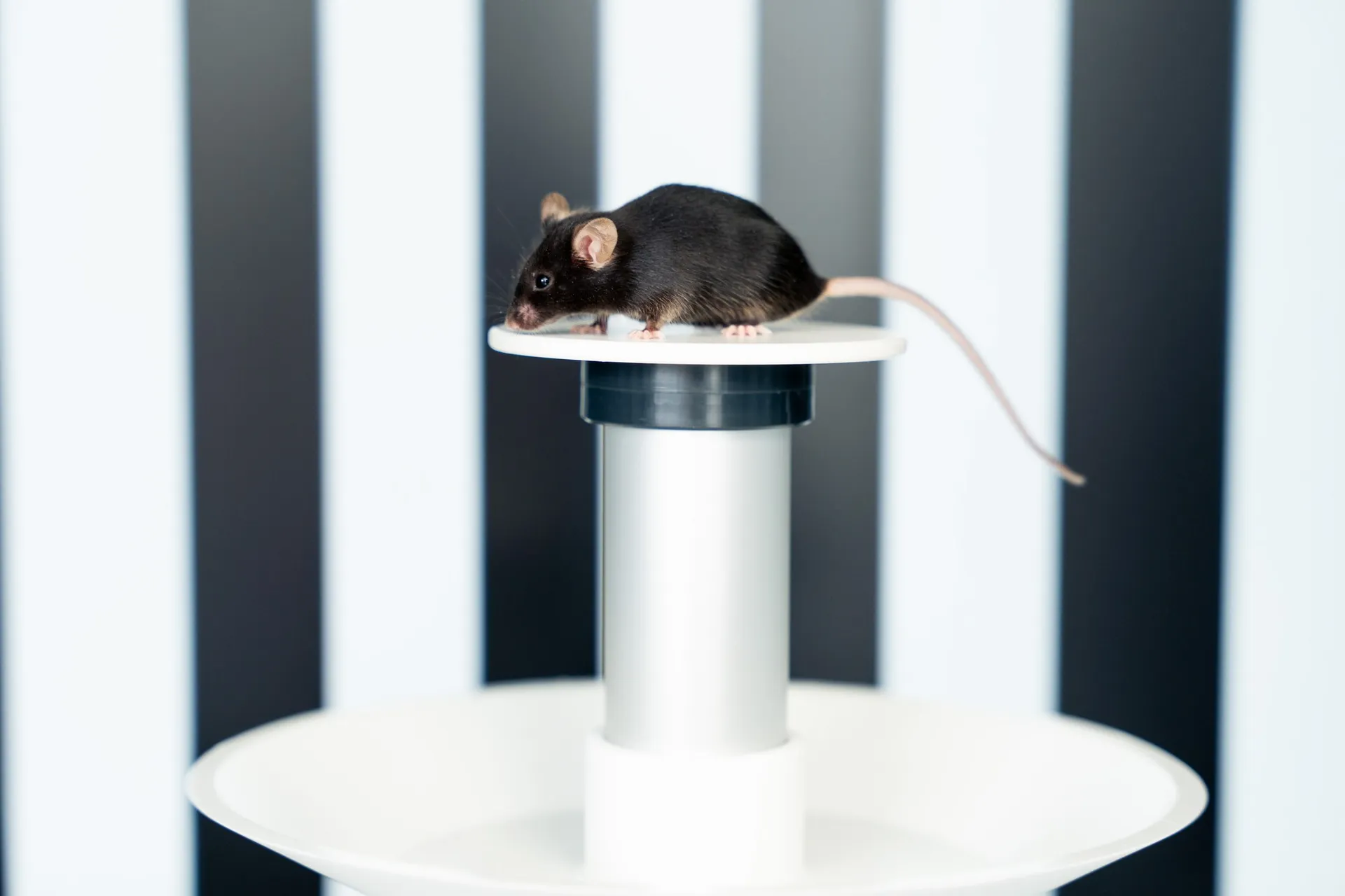
– Behavioral assessment (OMR), visual acuity and contrast sensitivity
– Functional assessment (fERG)
– Immunohistochemistry (IHC)
Highlights of this model
RP drug screening
Valuable tool to study retinal degeneration and to evaluate the efficacy of therapy candidates for RP
Non-invasive follow-up
Efficient, non-invasive model follow-up using our in vivo imaging and functional assessment technologies
Deep tissue insights
Detailed retinal analysis through immunohistochemistry and histology revealing the effects of therapy candidates
References
Receive model details
Interested to learn more? Fill out the form below and we will email you a white paper on the disease model. Your information will not be added to any mailing lists or used for marketing purposes.
"*" indicates required fields
We are here to help
Whether you have a question about our preclinical models, capabilities, pricing or anything else, our team is ready to answer all your inquiries.
Related services
Flash Electroretinography
Flash ERG is a non-invasive method to assess retinal function in preclinical eye disease models.
Learn moreOptomotor Reflex
Experimentica offers behavioral assessment and optomotor response testing to evaluate visual acuity and contrast sensitivity in rodent models.
Learn moreIn vivo imaging
Experimentica offers extensive in vivo imaging capabilities for high-resolution ocular assessments across species.
Learn moreCheck out our latest news and activities
All News