Modeling Diabetic Retinopathy in rodents
A brief overview of streptozotocin-induced diabetic retinopathy in rodents, highlighting strain-specific differences, quantitative assessment of retinal vascular leakage, identification of the most consistent and reliable readout, and animal welfare considerations.
Various inducible preclinical rodent models exist that recapitulate key aspects of diabetic retinopathy (DR)-related pathology. For instance, intravitreally administered vascular endothelial growth factor (VEGF) induces short-term vascular leakage, while an induction of choroidal neovascularization (CNV) with laser is great at evaluating anti-angiogenic effects. Similarly, oxygen-induced retinopathy (OIR), although closely modelling the retinopathy of prematurity, is also great at assessing retinal neovascularization. However, these models develop the pathology that is limited to a narrow time window, ranging from several hours (VEGF) to days (OIR) or a week (CNV). When lengthier follow-up is desired, the streptozotocin (STZ) model is widely recommended. Yet before exploring STZ-induced changes in more detail, consider this: even after 16-20 weeks, expect subtle microvascular alterations rather than dramatic remodeling.
Key readouts in Streptozotocin-Induced Diabetic Retinopathy
STZ is a naturally occurring toxin that selectively destroys insulin-producing beta cells in the pancreas. At Experimentica, we model DR in rodents using intraperitoneal injection of STZ. Chronic hyperglycemia, significant thinning of retinal layers, and impaired visual function are the most useful readouts.
Rat strain-dependent differences in STZ model
In 2018, we piloted a study with STZ-induced DR in three rat strains: Brown Norway, Long Evans and Wistar rats. All three rat strains were susceptible to significant and stable elevation of the blood glucose levels after STZ administration.
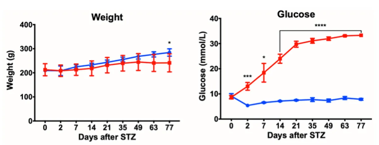
Fig. 1. Body weight and blood glucose level after STZ induction in Brown Norway rats
Vitreofluorophotometry, in vivo quantitative measurement of the inner blood-retinal barrier integrity, was used to detect inner retinal capillaries leakage. Both Brown Norway and Wistar rats showed increased retinal capillaries leakage starting at follow-up day 7 (Wistars) or day 21 (Brown Norways) after STZ treatment.
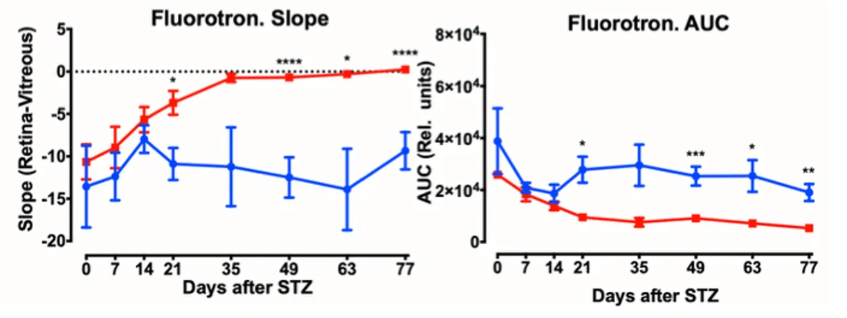
Fig. 2. Increased retinal vascular leak after STZ induction as assessed using vitreofluorophotometry in Brown Norway rats
Interestingly, Long Evans rats did not exhibit relevant DR-related changes despite successfully induced hyperglycemia. These data were presented at The European Association for Vision and Eye Research (EVER) conference in Nice, France.
A systematic review of diabetic retinopathy pathology in STZ model
To reveal the most consistent and reliable readout in the rodent STZ model, we conducted a systematic review of published articles. From 21 articles that met pre-defined inclusion criteria, a deterioration of visual function was reported in all as assessed either locally using electrophysiology or at the level of occipital cortex using visual evoked potentials. Our review paper was published in 2022 and is freely available for download.
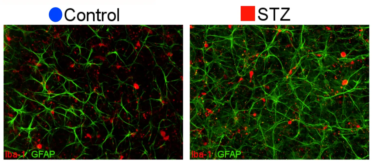
Fig. 3. Retinal flatmounts immunostained against microglial marker (Iba-1, in red) and astrocytic marker (GFAP, green) from healthy control and STZ-induced animal.
Electroretinography indicates a decline in photoreceptor and bipolar cell function in STZ-induced mice
In 2023, at The Association for Research in Vision and Ophthalmology (ARVO) annual meeting in New Orleans, LA, USA, we presented data that confirms previous findings of significant decrease of ERG parameters and retinal thickness after 70 day-long study in mice. The a- and b-wave amplitudes decreased by 45-50% relative to baseline values associated with a 7% reduction in inner retinal thickness.
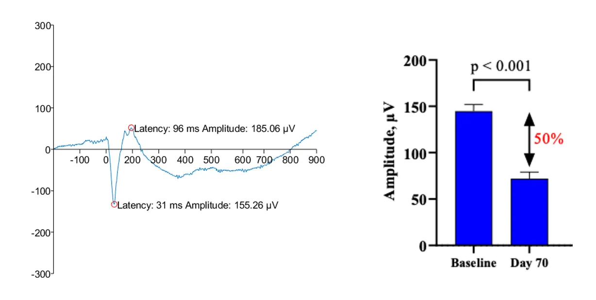
Fig. 4. A representative trace of fERG and the decrease of the a-wave amplitude 70 days after STZ induction in C57Bl/6J mice
Animal welfare in STZ studies
Our recent insights on STZ model are related to animal welfare. Recent evaluations of animal welfare complications in STZ-induced models demonstrated that Brown Norway rats exhibit higher susceptibility to adverse effects than Sprague Dawley rats. Our paper published in 2025 provides guidelines helping to adhere to the 3Rs principles.
For more information on diabetic retinopathy-related preclinical models, please fill the form or meet our scientific team at upcoming events.
Giedrius Kalesnykas
, PhD
Executive Chairman, Chief R&D Officer