ARVO 2025 presentation: Thioredoxin system and HIF1a pathway activation by oxidative stress in human iPSC-RPE cells
Scientific publications
04.05.2025
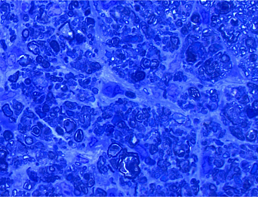
In vitro: Retinal pigment epithelium (RPE) cells exhibit a high susceptibility to reactive oxygen species (ROS) due to their elevated metabolic activity. The accumulation of ROS is a critical factor contributing to the pathogenesis of both dry and wet forms of age-related macular degeneration (AMD). The thioredoxin (TRX) system represents one of the two primary thiol-dependent antioxidant defense mechanisms in mammalian cells. The objective of this study was to investigate the concentration-dependent activation and response of the TRX system to ROS induced by RPE specific exogenous oxidative agent, sodium iodate (NaIO3).
Methods
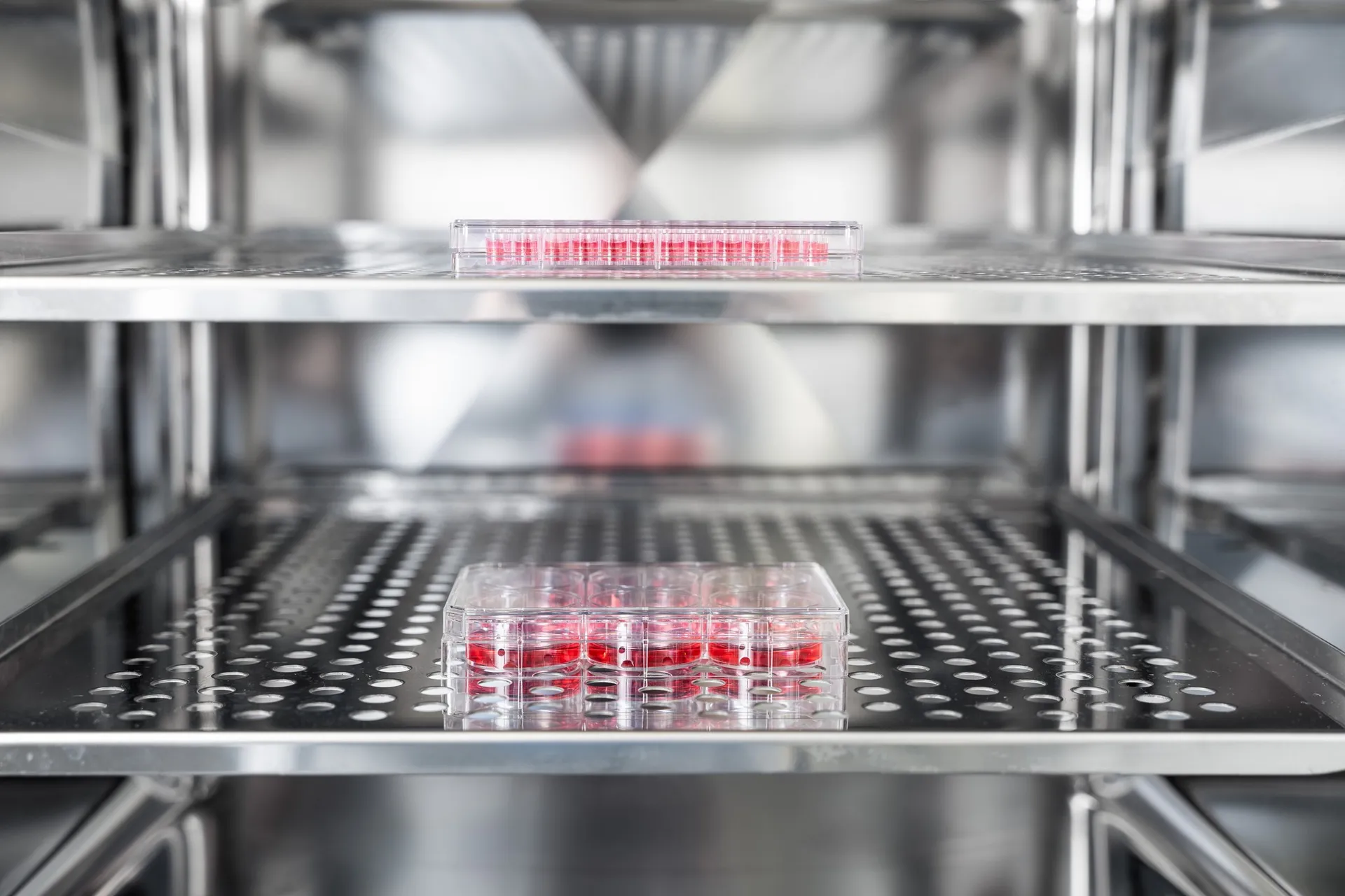
Human induced pluripotent stem cell derived RPE (iPSC-RPE) cells were cultured in 96-well plates. Oxidative stress was induced with NAIO3 (0-4 mM) for 24 h. Cell viability was assessed by using two different methods: resazurin cell viability assay and lactate dehydrogenase (LDH) release. Extracellular vascular endothelial growth factor A (VEGFA) levels were measured using ELISA. ROS production was assessed using a general ROS indicator. Cells were lysed in TRIzol, RNA was purified and converted to cDNA. RT-qPCR analysis included VEGFA, hypoxia inducible factor 1 subunit alpha (HIF1A), TRX, and TRX interacting protein (TXNIP) genes normalized to endogenous glyceraldehyde-3-phosphate dehydrogenase (GAPDH) control. qPCR data was analyzed with 2–ΔΔCT method for relative gene expression analysis.
Results
ROS levels significantly increased with low concentrations of NaIO3 (0-2 mM), peaking at 1 mM (7.7-fold higher compared to 0 mM), while viability indicators did not show decreased cell viability. ROS formation decreased at 1-4 mM, with a significant reduction (P < 0.001) at 4 mM compared to 1 mM. VEGFA release showed a significant increase at 1 mM (P = 0.02) and 4 mM (P < 0.001) compared to 0 mM. LDH release was significantly elevated (P < 0.001) at 3-4 mM compared to 0 mM. VEGFA, HIF1A, and TRX genes were significantly upregulated (P < 0.003) at 2-4 mM, and TXNIP gene was significantly downregulated (P < 0.05) at 2-4 mM.
Conclusions
NaIO3 significantly increased ROS formation in iPSC-RPE cells at low concentrations indicating intracellular oxidative stress damage without signs of decreased cell viability. Oxidative stress activated TRX system and TRX-HIF1α pathway, mitigating ROS formation and enhancing VEGFA release as part of the cellular response.
Download our poster
Authors
Robertas Cesna, Olga Vergun, Anni Kolehmainen, Dovilė Litvinavičiūtė, Giedrius Kalesnykas, Jenni J. Hakkarainen