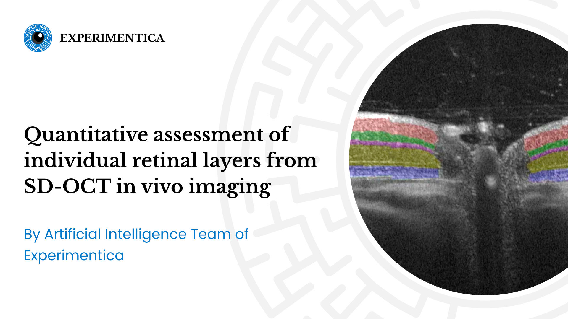
Merveilles de couleur dans la rétine - Experimentica designed an AI-based model capable of recognizing and quantifying retinal thickness layers from multiple SD-OCT scans.
Experimentica team designed an AI-based model, which is capable of automatically recognizing and quantitatively evaluating the thickness of individual retinal layers from multiple SD-OCT scans.
This clip from Experimentica’s YouTube channel shows an SD-OCT scan of the rat retina with nerve fiber layer together with retinal ganglion cell layer (first layer from the top, in grey), inner plexiform layer (in red), inner nuclear layer (in green), outer plexiform layer (in purple), outer nuclear layer (forest green), and outer segments of photoreceptor cells (blue).
Contact us to discuss how we integrate AI in our image analysis workflow for your studies.
All our facilities are AAALAC accredited, reflecting our commitment to the highest standards of animal care in research.
Copyright: Experimentica Ltd. 2026
Manage Consent
To provide the best experiences, we use technologies like cookies to store and/or access device information. Consenting to these technologies will allow us to process data such as browsing behavior or unique IDs on this site. Not consenting or withdrawing consent, may adversely affect certain features and functions.
Functional
Always active
The technical storage or access is strictly necessary for the legitimate purpose of enabling the use of a specific service explicitly requested by the subscriber or user, or for the sole purpose of carrying out the transmission of a communication over an electronic communications network.
Preferences
The technical storage or access is necessary for the legitimate purpose of storing preferences that are not requested by the subscriber or user.
Statistics
The technical storage or access that is used exclusively for statistical purposes.The technical storage or access that is used exclusively for anonymous statistical purposes. Without a subpoena, voluntary compliance on the part of your Internet Service Provider, or additional records from a third party, information stored or retrieved for this purpose alone cannot usually be used to identify you.
Marketing
The technical storage or access is required to create user profiles to send advertising, or to track the user on a website or across several websites for similar marketing purposes.