AI for image analysis
AI-driven image analysis for in vivo studies, delivering faster, unbiased results and deeper insights for your preclinical ocular programs.
Learn more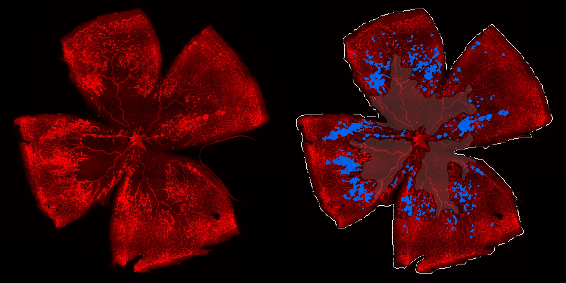
AI-driven image analysis for in vivo studies, delivering faster, unbiased results and deeper insights for your preclinical ocular programs.
Learn more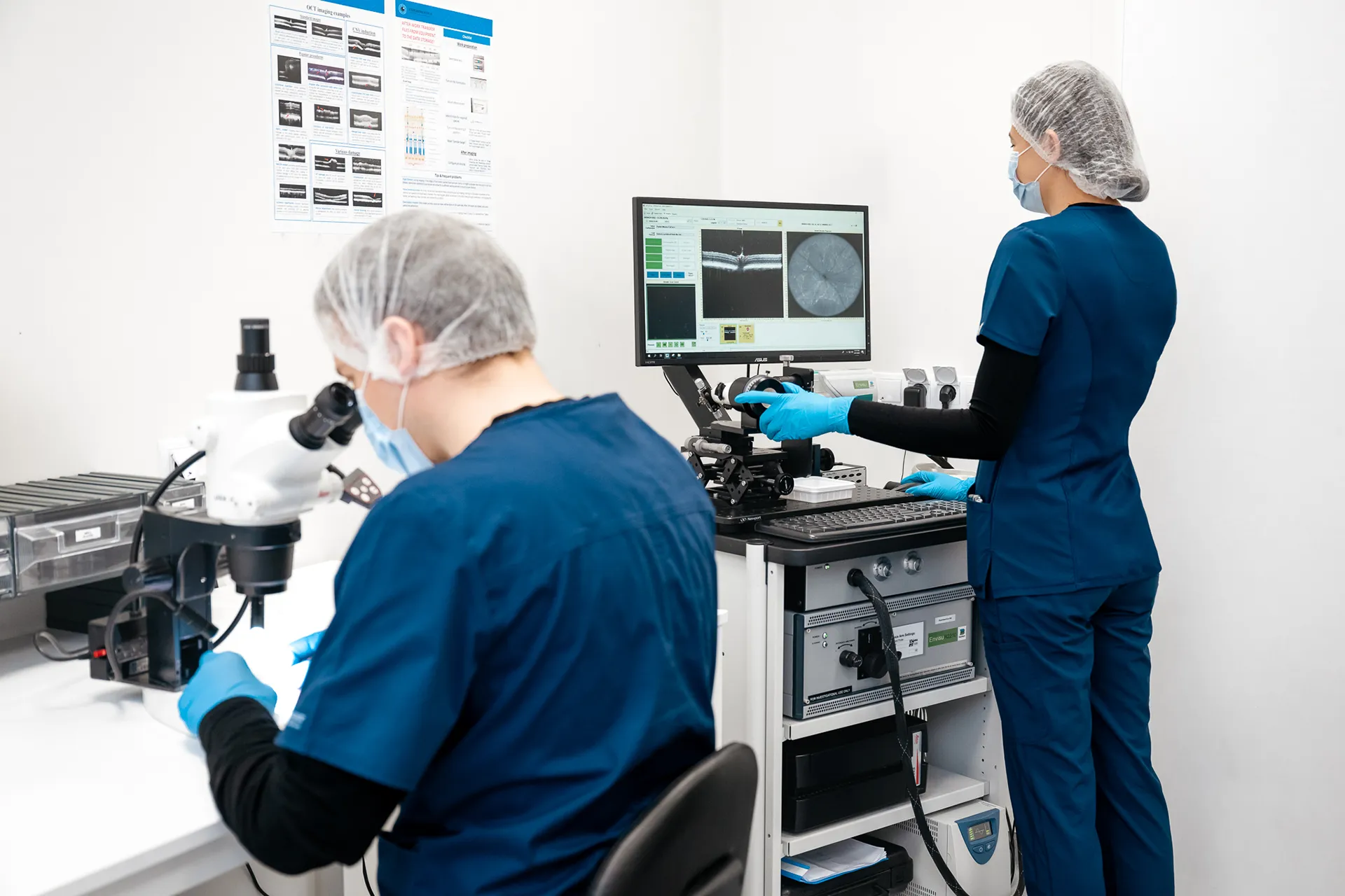
Experimentica offers extensive in vivo imaging capabilities for high-resolution ocular assessments across species.
Learn more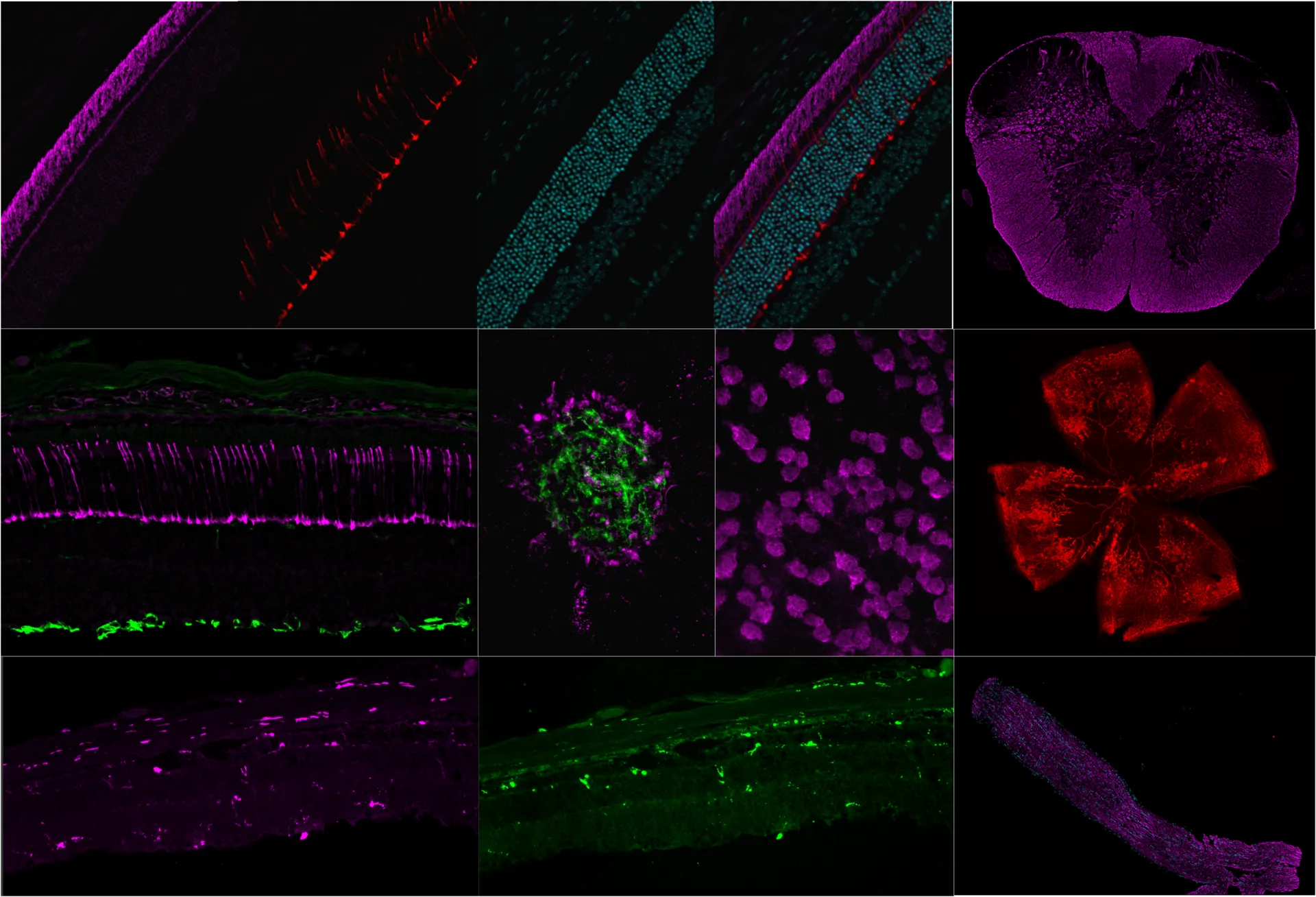
Experimentica offers a wide range of single and multiplex immunofluorescence labeling to explore disease pathogenesis and therapeutic targets.
Learn more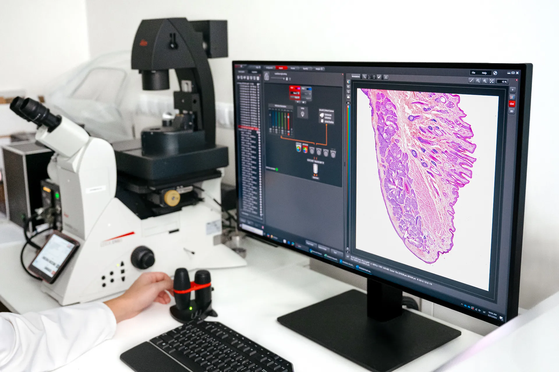
Histological staining techniques for ocular and nervous system tissues to support detailed analysis.
Learn more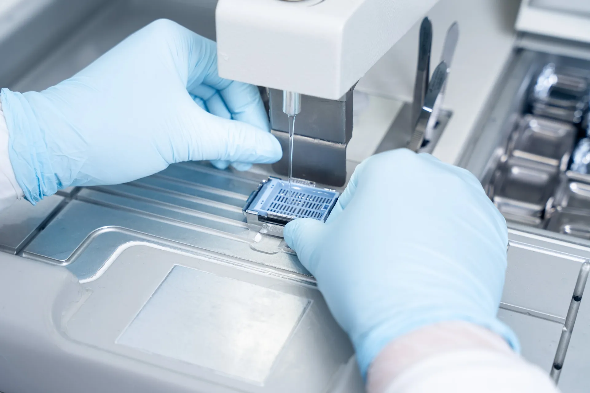
Specialized processing of ocular and neural tissues employing methods such as flat mounting, paraffin and cryo embedding, and precision sectioning.
Learn more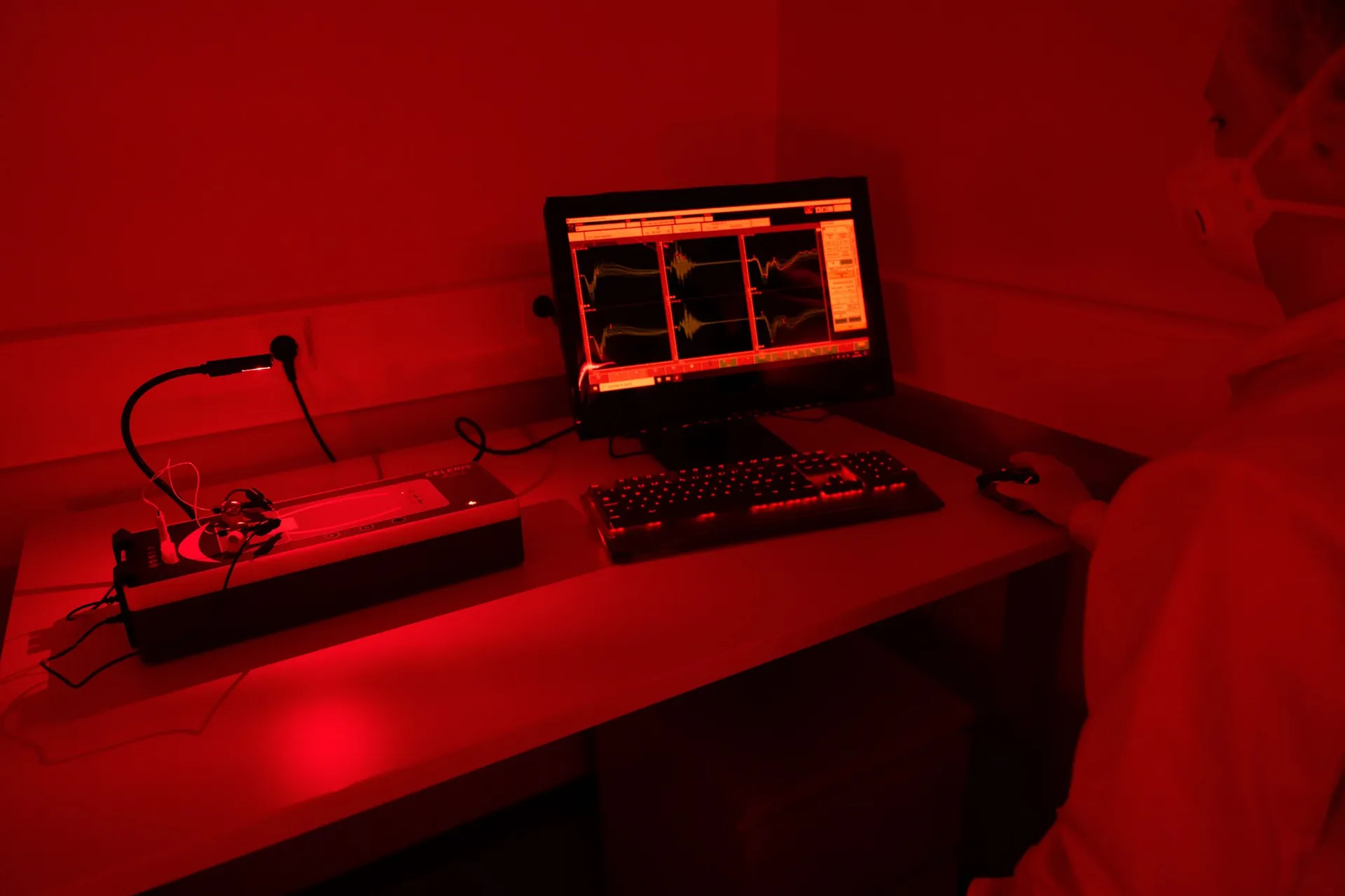
Flash ERG is a non-invasive method to assess retinal function in preclinical eye disease models.
Learn more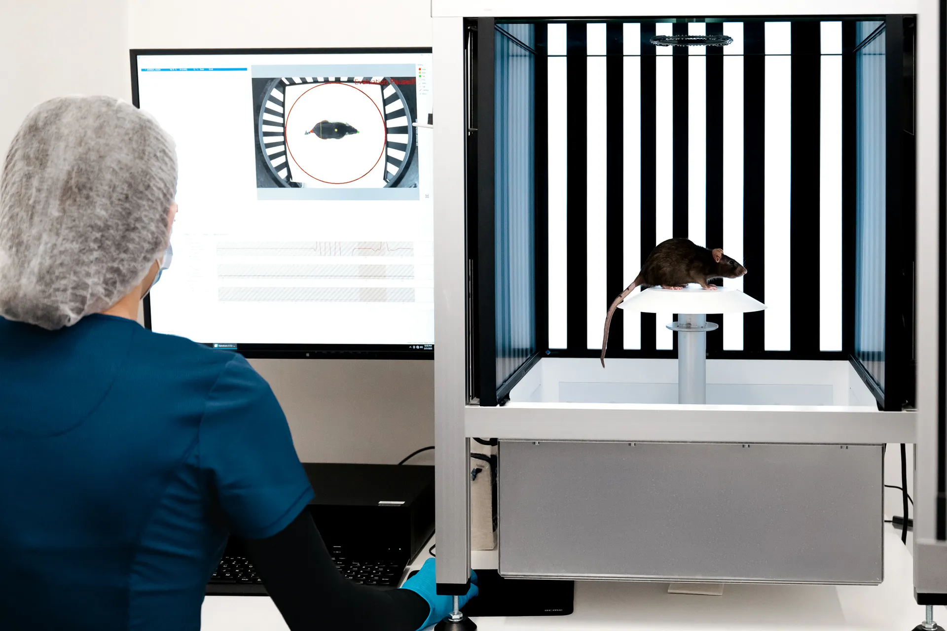
Experimentica offers behavioral assessment and optomotor response testing to evaluate visual acuity and contrast sensitivity in rodent models.
Learn more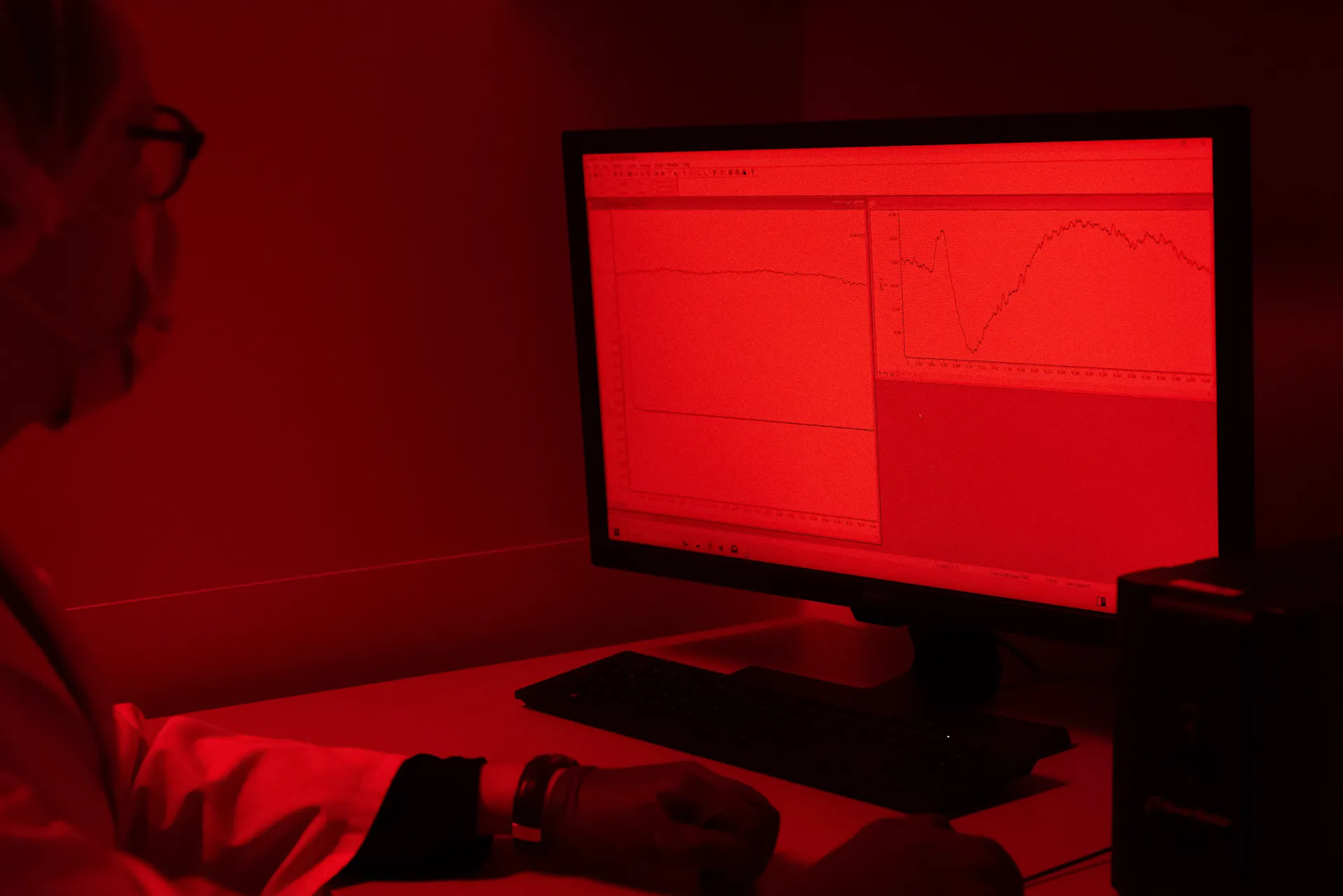
Pattern ERG is a non-invasive method to assess retinal ganglion cell function and monitor dysfunction in preclinical eye disease models.
Learn more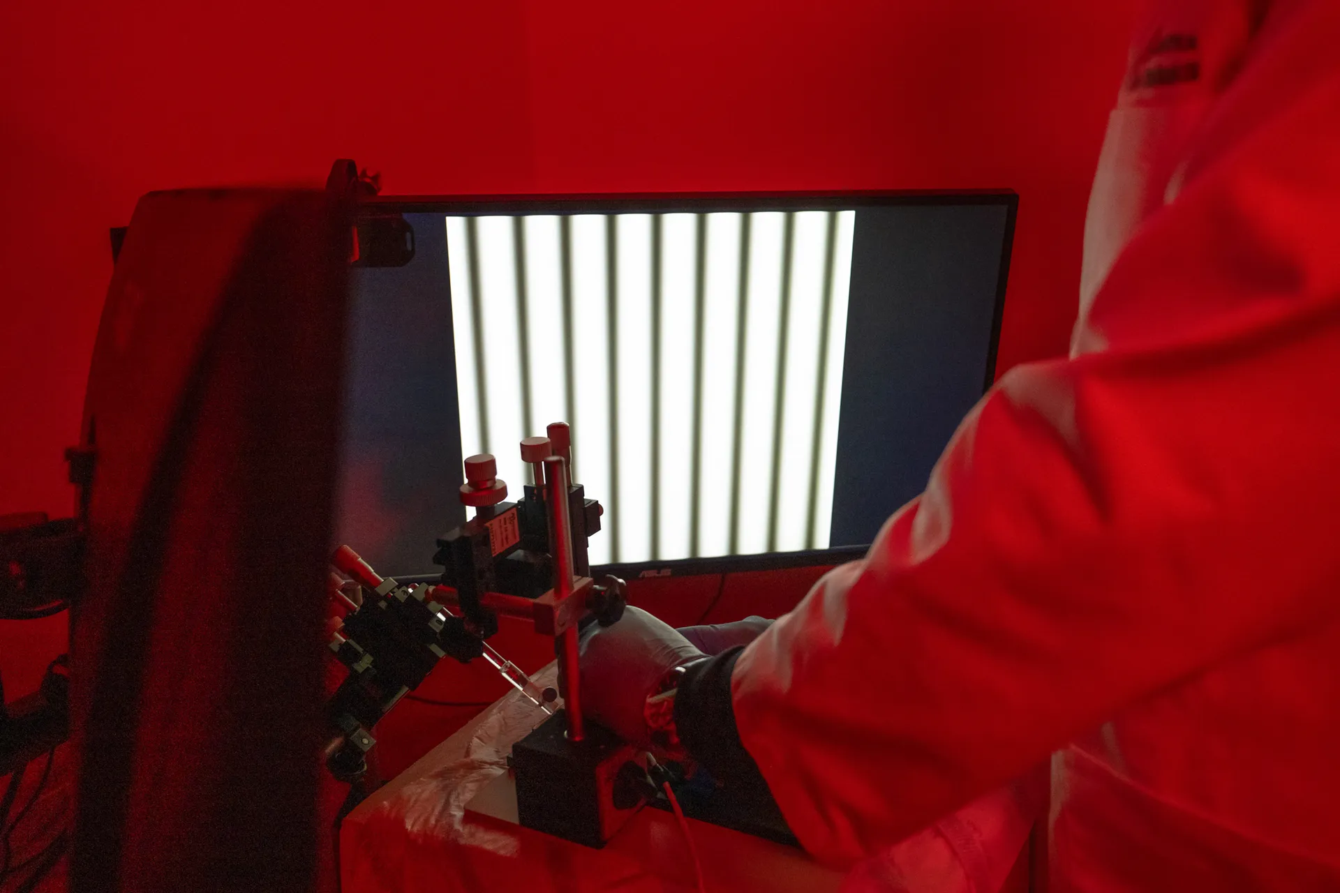
Visual evoked potential recording is used to assess visual pathway function from retina to cortex in models of glaucoma and optic neuropathies.
Learn more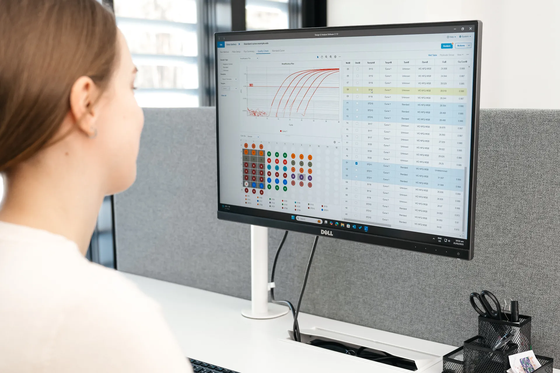
qPCR measures gene expression in ocular tissues, supporting disease research and treatment evaluation.
Learn moreAll our facilities are AAALAC accredited, reflecting our commitment to the highest standards of animal care in research.
Copyright: Experimentica Ltd. 2026