Our expertise
Histological staining
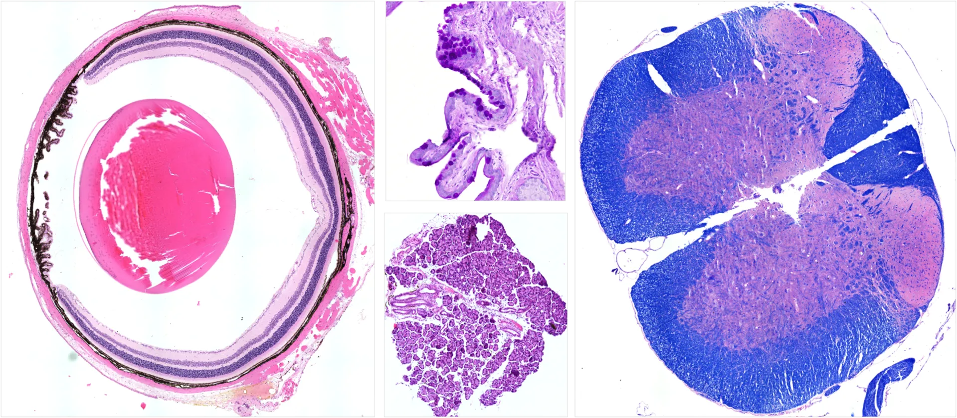
Accurate tissue characterization is crucial in ocular pathology. Our team provides a comprehensive range of classical and specialized histological stains designed to unveil the intricate structural and cellular details of the eye. Whether working with cryosections, paraffin-embedded, or resin-embedded tissues, we ensure tissues have optimal preservation and visualization for ocular morphology assessment.
Hematoxylin and Eosin (H&E) staining
Hematoxylin and Eosin (H&E) staining, the gold standard in histopathology, offers clear contrast between cell nuclei and cytoplasmic or extracellular structures. It is an ideal tool for assessing general tissue architecture, detecting inflammation, and identifying degenerative or neoplastic changes in ocular tissues.
Periodic Acid–Schiff (PAS) staining
Periodic Acid–Schiff (PAS) staining highlights carbohydrates and mucopolysaccharides, enabling precise visualization of basement membranes, glycogen, and other polysaccharide-rich structures. In the eye, it is particularly useful for studying the corneal epithelium, conjunctiva, and retinal layers.
Luxon Fast Blue (LFB)
Luxol Fast Blue (LFB) is the preferred stain for myelin, making it invaluable in assessing optic nerve integrity and demyelinating processes. When combined with counterstains, LFB provides detailed insights into the relationship between neural fibers and supporting glial elements.
In addition to these specialized stains, we offer a wide selection of other histochemical techniques for detecting connective tissue components, elastic fibers, pigments, and lipids. Our staining services are optimized for reproducibility, clarity, and compatibility with a range of downstream analyses, from light microscopy to advanced image quantification.
We are here to help
Whether you have a question about our preclinical models, capabilities, pricing or anything else, our team is ready to answer all your inquiries.
Related services
Tissue processing
Specialized processing of ocular and neural tissues employing methods such as flat mounting, paraffin and cryo embedding, and precision sectioning.
Learn moreImmunohistochemistry
Experimentica offers a wide range of single and multiplex immunofluorescence labeling to explore disease pathogenesis and therapeutic targets.
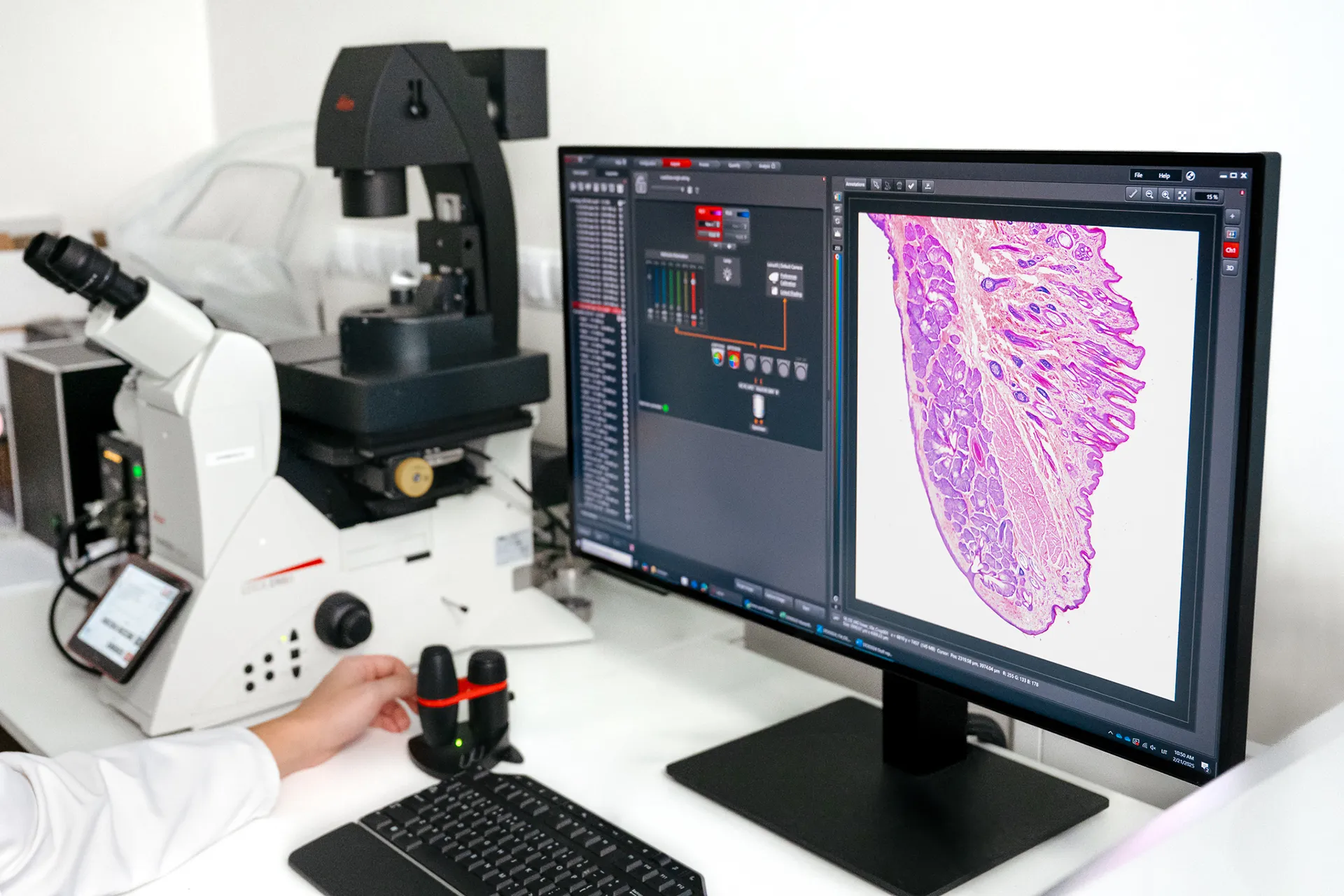
Learn moreAI for image analysis
AI-driven image analysis for in vivo studies, delivering faster, unbiased results and deeper insights for your preclinical ocular programs.
Learn moreCheck out our latest news and activities
All News