


Summary: RPE cell monolayers make a tight paracellular barrier that can be used to screen early-stage drug candidates.
Model Description
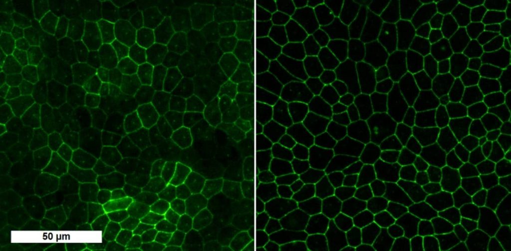
Retinal pigment epithelium (RPE) is a tight cell monolayer between retina and choroidal blood. RPE form the outer blood-retinal barrier (oBRB) restricting the drug entry to retina after systemic administration. PCi-RPE1426 cell monolayers represent tight paracellular barrier, thus, this in vitro cell model can be used to screen drug molecules in early phase of drug development.
| Species | RPE cells derived from human induced pluripotent stem cells (PCi-RPE1426, Phenocell, France) |
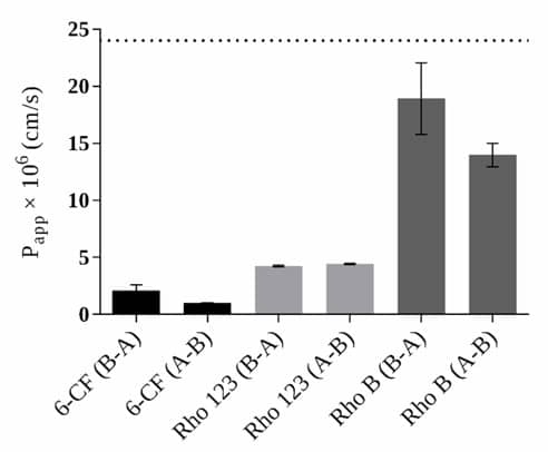
| Assessment | The permeability experiments across PCi-RPE1426 cell monolayers are conducted in Transwell® cell culture inserts (Corning Inc., Corning, NY, USA) in basolateral-to-apical direction (B-A, represent the transport from the choroid to the retina) to measure the transport of study molecules and standard molecules. The cumulative amount of study molecules in the receiver chamber versus time is measured. |
| Standard molecules | Low permeability standard: 6-carboxyfluorescein High permeability standard: Rhodamine B |
| Read-outs | The rate (apparent permeability coefficient, Papp) of the study molecules is compared to the Papp values of the low and high permeability markers. |
Outcomes and Read-Outs
Imaging
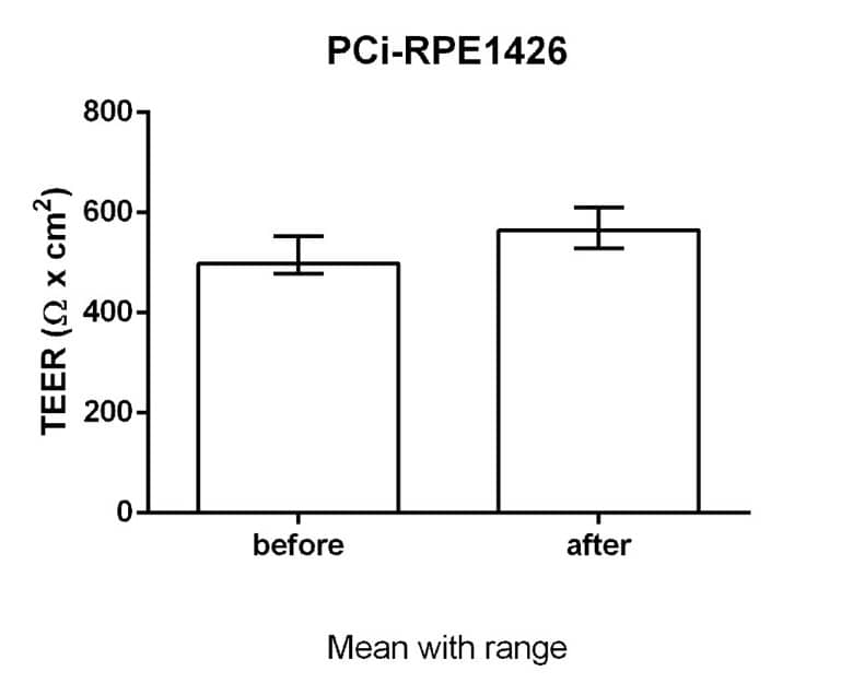
Permeability measurements
References
- Maruotti J, Wahlin K, Gorrell D, Bhutto I, Lutty G, Zack DJ. A simple and scalable process for the differentiation of retinal pigment epithelium from human pluripotent stem cells. Stem Cells Transl Med. 2013,2(5):341-354. doi: 10.5966/sctm.2012-0106.
- Hakkarainen JJ, Maruotti J, Seppänen A, Onteniente B, Kalesnykas G, Reinisalo M. hiPSC-derived RPE cells: Characterization of blood-retinal barrier properties and drug permeability. ARVO2016 Poster. Investigative Ophthalmology & Visual Science September 2016, Vol.57, 268.