Summary: This year the work of Experimentica's scientists at the forefront of Ophthalmology research will be featured at the Neuroscience 2021.

Content
The Society for Neuroscience is the world’s largest organization of scientists and physicians devoted to understanding the brain and the nervous system. This year the work of Experimentica’s scientists at the forefront of Ophthalmology research will be featured at the Neuroscience 2021.
Featured presentations
Our team will present the three following abstracts during the conference. Posters will be available for download after the conference.
A B S T R A C T
Cultured hiPSC-RPE cells exhibit increased resistance to oxidative stress triggers throughout differentiation and maturation
November 9, 2021, 10:45am CST
Presenting: Anita Ghosh
Human induced pluripotent stem cell-derived retinal pigment epithelium (hiPSC-RPE) cells more closely exhibit the human RPE than existing RPE-derived cell lines, making them a promising tool for drug discovery and development for ophthalmic conditions affecting the RPE, including age-related macular degeneration and geographic atrophy. However, there is a lack of rigorous scientific data regarding the response of hiPSC-RPE cells to oxidative stress triggers. The purpose of this study was to determine the susceptibility of hiPSC-RPE cells to exogenously applied oxidative stress (tert-butyl hydroperoxide; tBHP) and an oxidizing agent (sodium iodate; NaIO3) that triggers intracellular oxidative stress and to investigate their effects on hiPSC-RPE differentiation and maturation.
HiPSC-RPE cells were seeded on Matrigel®-coated 96-well cell culture plates and differentiated according to the manufacturer’s instructions. After varying amounts of differentiation time in vitro (days in vitro, DIV), cells were challenged with either tBHP (0.1 to 1.0 mM) or NaIO3 (0 to 18 mM). Cell viability was assessed by resazurin assay and lactate dehydrogenase (LDH) release assay. Reactive oxygen species (ROS) generation was quantified by chloromethyl 2′,7′-dichlorodihydrofluorescein diacetate (CM-H2DCFDA) assay and vascular endothelial growth factor (VEGF) release was quantified by ELISA.
HiPSC-RPE cells exhibited a dose-response to increasing doses of tBHP and NaIO3. In tBHP experiments, IC50 values for tBHP from resazurin assay were ~0.4 mM from DIV13 to DIV19, ~0.7 at DIV26 and ~0.9 mM at DIV28. LDH release showed a similar shift (EC50 values for tBHP: range 0.4 mM to 0.9 mM). ROS generation was significantly attenuated at DIV26 and DIV28, which was positively associated with VEGF release.
Similarly, NaIO3 induced a dose-response in the cells with IC50 (resazurin assay) and EC50 (LDH assay) values of 7.8 mM. NaIO3 elicited a significant 26-fold increase in ROS generation at the lowest dose of 2 mM NaIO3, which slowly decreased with higher doses, likely attributable to a dose-dependent toxicity.
Our data suggest that hiPSC-RPE cells continue to mature over time in vitro, manifesting as increasing resistance to exogenously-applied oxidative stress. Differences in the correlation between insult (tBHP vs. NaIO3) and cell viability are likely due to mechanistic differences in the generation of intracellular vs. exogenously-applied oxidative stress. HiPSC-RPE cells exhibit a strong endogenous antioxidant system and potent VEGF response, making them a useful model for in vitro applications.
A B S T R A C T
Functional effects of astrocyte-derived exosomes on neurite outgrowth
November 10, 2021, 9:30am CST
Presenting: Simon Kaja
Exfoliation glaucoma is the most common identifiable secondary form of open-angle glaucoma and associated with deposition of extracellular fibrillar material in ocular tissues.
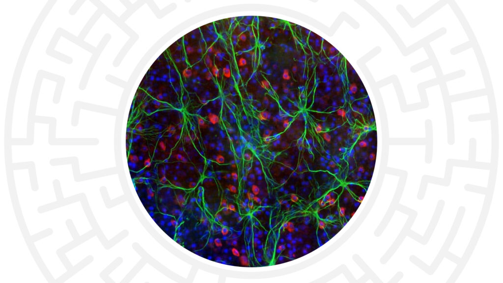
Genetic polymorphisms in lysyl oxidase-like 1 (Loxl1) are the most common cause of exfoliation glaucoma. The objective of this study was to determine the functional effects of extracellular vesicles (exosomes) derived from Loxl1-deficient primary optic nerve head astrocytes (ONHAs) on neuronal differentiation and viability. We generated an ONHA culture with ~50% Loxl1 knockdown by transducing ONHAs with lentivirus harboring Loxl1- targeting shRNA. Exosomes from primary cultures of rat ONHAs (naïve, scramble- and Loxl1 shRNA-transduced) were prepared by ultracentrifugation. Exosomes were characterized by nanoparticle tracking analysis and immunoblotting. Uptake of exosomes into rat E18 primary cortical neurons was assessed by quantitative microscopy. The functional effects of exosomes on primary neurons in vitro were determined by quantification of cell viability and neurite outgrowth after addition of exosomes. ONHA-derived exosomes expressed the prototypical markers, Alix and CD63, but were devoid of CD81. Exosomes derived from naïve as well as scramble- and Loxl1 shRNA-transduced ONHAs were similar in size (119.8 ± 4.3, 121.9 ± 2.8 and 121.1 ± 2.5 nm, respectively, n = 3, P = 0.89). Uptake of exosomes into primary neurons was time-dependent, peaking at 4h after addition (n = 3, P < 0.05).
Viability of rat primary E18 cortical neuronal cultures did not differ in the presence of exosomes. However, naïve and scramble-transduced ONHA exosomes increased neurite outgrowth of primary cortical neurons by 20.1 ± 3.3% and 18.7 ± 2.0%, respectively (n=5, P < 0.05 compared with media alone). In contrast, exosomes from Loxl1 shRNA-transduced ONHAs prevented any increase in neurite outgrowth (2.2 ± 4.0% change; n = 5, P = 0.98).These data suggest ONHA-derived exosomes can exert functional effects on primary neurons in vitro. Altered exosome-mediated neuron-glia signaling in exfoliation glaucoma characterized by a chronic reduction of LOXL1 expression may contribute to the ocular manifestations of the disease.
A B S T R A C T
Structure-function relationships in the Brown Norway rat streptozotocin-induced diabetic retinopathy model
November 11, 2021, 11:00am CST
Presenting: Inesa Lelyte
The rat streptozotocin-induced model for diabetic retinopathy (DR) remains the most-commonly used experimental paradigm for drug discovery, as it recapitulates many of the complex phenotypes seen in patients. Advances in in vivo ocular imaging provide an opportunity to study early pathological processes in this model and to establish structure-function relationships.
We recently performed a systematic literature review that included 662 records of preclinical STZ studies from the past 10 years. Of those, only one study was performed in pigmented rats, specifically Brown Norway (BN) rats. The BN strain is widely used in ocular drug discovery and the presence of pigmentation allows for the detection of potential confounding pharmacologic effects due to melanin binding. This highlights an urgent need for structure-function analysis of DR phenotypes in BN rats.
BN rats (male, 3-4 months old) received two IP injections of STZ (65 mg/kg in citrate buffer, pH 4.5). Glucose levels and body weight were monitored twice per week. Rats were followed in vivo for 8-12 weeks by vitreofluorophotometry (VFP; OcuMetrics, Inc.), flash electroretinography (fERG; Diagnosys LLC), and spectral domain-optical coherence tomography (SD-OCT; Leica Microsystems, Inc.). Ocular tissues were collected for quantification of acellular capillaries and pericyte loss using trypsin digested retinal whole mounts, Evanʼ’s Blue (EB) extravasation assay and biomarker analysis. Majority of the rats developed hyperglycemia (>16 mmol/l) week 1 post-STZ (p < 0.001). Hyperglycemic rats presented significantly reduced body weight gain compared to the naïve rats by day 7 (p < 0.001). VFP showed mild progressive retinal leakage by week 3 post-STZ, which was statistically significant by week 5 (p < 0.01). fERG showed decreased photoreceptor responses by week 6 with increased latencies (p < 0.05). Inner retinal thickness measured by SD-OCT decreased by week 8-9 (p < 0.001). Pericyte ghosts increased significantly in diabetic animals compared to naïve rats (p < 0.05). EB levels in the retina showed a statistically significant increase, suggesting presence of blood-retinal barrier rupture.
Our data demonstrate that the BN strain reproduces the known hallmarks of diabetic retinopathy, as seen in albino rat strains. Detailed analysis of the structure-function relationship in the BN rats reveal changes consistent with significant progressive pro-angiogenic vascular changes and retinal degeneration starting as early as 4 weeks post-STZ induction. The pigmented BN STZ model for DR offers unique opportunities for drug discovery.