In vivo models
Two-Stage Laser-Induced Subretinal Fibrosis
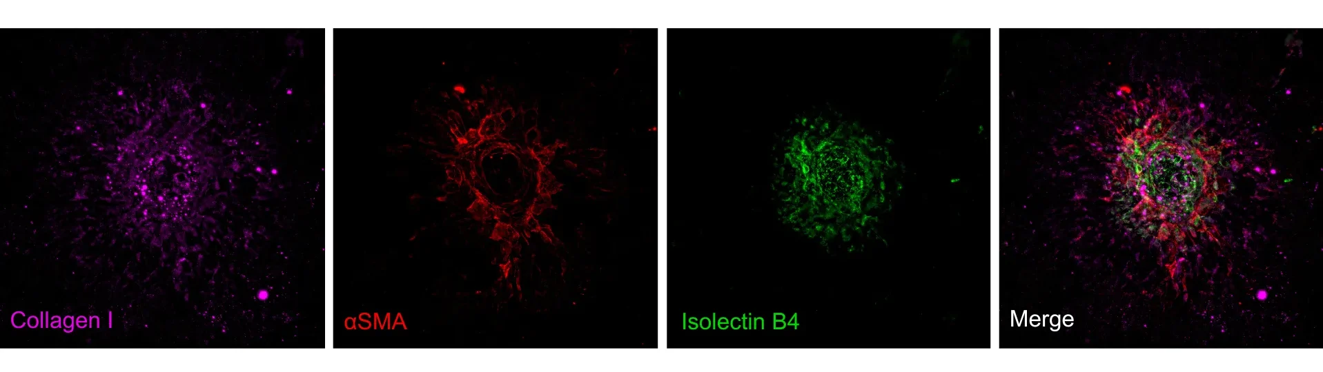
In 2020, Little and colleagues presented a two-stage laser-induced mouse model of subretinal fibrosis (Little et al., 2020), modifying the classical single-laser choroidal neovascularization (CNV) (Tobe et al., 1998). In the first stage, laser burns rupture Bruch’s membrane at multiple spots per eye, inducing CNV that typically peaks around Day 5 – Day 7 and begins to regress within three weeks. Before this regression occurs, at Day 7 a second laser burn is applied to each of the original lesions. This second lasering triggers renewed vascular leakage and, mild hemorrhage in a subset of lesions, converting the CNV into a stable fibrovascular membrane.
The resulting lesions recapitulate three hallmark pathological features of neovascular age-related macular degeneration (nAMD): persistent neovascularization, macrophage infiltration, and subretinal fibrosis. Because of this fibrovascular, non-regressing phenotype, the two-stage laser model is a valuable translational tool for dissecting the mechanisms of subretinal fibrosis and for testing the efficacy of anti-fibrotic treatments in nAMD.
Technical details
Wet AMD
Mouse
Diode laser
Topical (e.g. eye drops), intravitreal, subretinal, subconjuctival, systemic
In vivo imaging:
– Fluorescein angiography (FA)
– Spectral-domain optical coherence tomography (SD-OCT)
Morphological assessment:
– Histology
– Immunohistochemistry
Molecular biology:
– qPCR
– ELISA
– Western blotting
– In situ hybridization/RNAscope
References
- Ragauskas S, Kielczewski E, Vance J, Kaja S, Kalesnykas G. In Vivo Multimodal Imaging and Analysis of Mouse Laser-Induced Choroidal Neovascularization Model. JoVE 2018; 131.
- Dobi, E. T., Puliafito, C. A., & Destro, M. A new model of experimental choroidal neovascularization in the rat. Arch. Ophthalmol. Chic. Ill 1960. 107, 264-269 (1989).
Receive model details
Interested to learn more? Fill out the form below and we will email you a white paper on the disease model. Your information will not be added to any mailing lists or used for marketing purposes.
"*" indicates required fields
We are here to help
Whether you have a question about our preclinical models, capabilities, pricing or anything else, our team is ready to answer all your inquiries.

Related services
In vivo imaging
Experimentica offers extensive in vivo imaging capabilities for high-resolution ocular assessments across species.
Learn moreHistological staining
Histological staining techniques for ocular and nervous system tissues to support detailed analysis.
Learn moreImmunohistochemistry
Experimentica offers a wide range of single and multiplex immunofluorescence labeling to explore disease pathogenesis and therapeutic targets.
Learn moreWestern Blotting
Western blot detects protein expression and modifications in ocular tissues with high sensitivity and precision
Learn moreELISA
ELISA enables sensitive protein detection and quantification in ocular tissues and biofluids.
Learn moreRT-qPCR
qPCR measures gene expression in ocular tissues, supporting disease research and treatment evaluation.
Learn moreCheck out our latest news and activities
All News